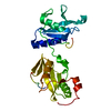
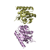
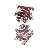
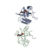
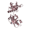
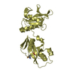
[English] 日本語
Yorodumi
- PDB-4ey0: Structure of tandem SH2 domains from PLCgamma1 -

+
Open data


ID or keywords:

Loading...

-
Basic information

Entry
Database: PDB / ID: 4ey0
TitleStructure of tandem SH2 domains from PLCgamma1
Components1-phosphatidylinositol 4,5-bisphosphate phosphodiesterase gamma-1
KeywordsHYDROLASE / SH2 Domain / PLCgamma specific array / Interaction Domain / fibroblast growth factor receptor 1
Function / homology
Function and homology information


Activated NTRK3 signals through PLCG1 / Activated NTRK2 signals through PLCG1 / calcium-dependent phospholipase C activity / Role of second messengers in netrin-1 signaling / PLC-gamma1 signalling / phosphoinositide phospholipase C / DAG and IP3 signaling / phospholipid catabolic process / phosphatidylinositol metabolic process / Phospholipase C-mediated cascade; FGFR3 ...Activated NTRK3 signals through PLCG1 / Activated NTRK2 signals through PLCG1 / calcium-dependent phospholipase C activity / Role of second messengers in netrin-1 signaling / PLC-gamma1 signalling / phosphoinositide phospholipase C / DAG and IP3 signaling / phospholipid catabolic process / phosphatidylinositol metabolic process / Phospholipase C-mediated cascade; FGFR3 / Phospholipase C-mediated cascade; FGFR2 / phosphatidylinositol phospholipase C activity / Phospholipase C-mediated cascade; FGFR4 / Erythropoietin activates Phospholipase C gamma (PLCG) / COP9 signalosome / Phospholipase C-mediated cascade: FGFR1 / phospholipase C activity / neurotrophin TRKA receptor binding / positive regulation of endothelial cell apoptotic process / phosphatidylinositol-mediated signaling / EGFR interacts with phospholipase C-gamma / positive regulation of vascular endothelial cell proliferation / PLCG1 events in ERBB2 signaling / Signaling by ALK / positive regulation of epithelial cell migration / Synthesis of IP3 and IP4 in the cytosol / Fc-epsilon receptor signaling pathway / PECAM1 interactions / Generation of second messenger molecules / Signaling by ALK fusions and activated point mutants / RET signaling / glutamate receptor binding / positive regulation of blood vessel endothelial cell migration / Role of phospholipids in phagocytosis / Signaling by FGFR4 in disease / release of sequestered calcium ion into cytosol / negative regulation of inflammatory response to antigenic stimulus / Signaling by FGFR3 in disease / cellular response to epidermal growth factor stimulus / ruffle / Signaling by FGFR2 in disease / Signaling by FGFR1 in disease / Downstream signal transduction / FCERI mediated Ca+2 mobilization / guanyl-nucleotide exchange factor activity / FCGR3A-mediated IL10 synthesis / positive regulation of release of sequestered calcium ion into cytosol / VEGFR2 mediated cell proliferation / cell projection / calcium-mediated signaling / FCERI mediated MAPK activation / Signaling by ERBB2 TMD/JMD mutants / modulation of chemical synaptic transmission / Schaffer collateral - CA1 synapse / Constitutive Signaling by EGFRvIII / Signaling by ERBB2 ECD mutants / epidermal growth factor receptor signaling pathway / Signaling by ERBB2 KD Mutants / receptor tyrosine kinase binding / ISG15 antiviral mechanism / ruffle membrane / positive regulation of angiogenesis / cell-cell junction / cell migration / Constitutive Signaling by Ligand-Responsive EGFR Cancer Variants / DAP12 signaling / lamellipodium / T cell receptor signaling pathway / in utero embryonic development / glutamatergic synapse / calcium ion binding / protein kinase binding / plasma membrane / cytosol / cytoplasm
Similarity search - Function
1-phosphatidylinositol 4,5-bisphosphate phosphodiesterase gamma-1, SH3 domain / Phosphatidylinositol-4, 5-bisphosphate phosphodiesterase gamma / PLC-gamma, C-terminal SH2 domain / PLC-gamma, N-terminal SH2 domain / Phosphoinositide phospholipase C family / Phospholipase C, phosphatidylinositol-specific, Y domain / Phosphatidylinositol-specific phospholipase C, Y domain / Phosphatidylinositol-specific phospholipase Y-box domain profile. / Phospholipase C, catalytic domain (part); domain Y / Phosphatidylinositol-specific phospholipase X-box domain profile. ...1-phosphatidylinositol 4,5-bisphosphate phosphodiesterase gamma-1, SH3 domain / Phosphatidylinositol-4, 5-bisphosphate phosphodiesterase gamma / PLC-gamma, C-terminal SH2 domain / PLC-gamma, N-terminal SH2 domain / Phosphoinositide phospholipase C family / Phospholipase C, phosphatidylinositol-specific, Y domain / Phosphatidylinositol-specific phospholipase C, Y domain / Phosphatidylinositol-specific phospholipase Y-box domain profile. / Phospholipase C, catalytic domain (part); domain Y / Phosphatidylinositol-specific phospholipase X-box domain profile. / Phosphatidylinositol-specific phospholipase C, X domain / Phosphatidylinositol-specific phospholipase C, X domain / Phospholipase C, catalytic domain (part); domain X / PLC-like phosphodiesterase, TIM beta/alpha-barrel domain superfamily / SH2 domain / C2 domain / Protein kinase C conserved region 2 (CalB) / SHC Adaptor Protein / C2 domain / C2 domain profile. / PH domain / C2 domain superfamily / PH domain profile. / Pleckstrin homology domain. / Pleckstrin homology domain / SH3 domain / SH2 domain / Src homology 2 (SH2) domain profile. / Src homology 2 domains / SH2 domain / Src homology 3 domains / EF-Hand 1, calcium-binding site / EF-hand calcium-binding domain. / SH2 domain superfamily / EF-hand calcium-binding domain profile. / SH3-like domain superfamily / Src homology 3 (SH3) domain profile. / SH3 domain / EF-hand domain / EF-hand domain pair / PH-like domain superfamily / 2-Layer Sandwich / Alpha Beta
Similarity search - Domain/homology
1-phosphatidylinositol 4,5-bisphosphate phosphodiesterase gamma-1
Similarity search - Component
Biological speciesHomo sapiens (human)
MethodX-RAY DIFFRACTION / SYNCHROTRON / MOLECULAR REPLACEMENT / Resolution: 2.8 Å
AuthorsCole, A.R. / Mas-Droux, C.P. / Bunney, T.D. / Katan, M.
CitationJournal: Structure / Year: 2012
Title: Structural and Functional Integration of the PLCgamma Interaction Domains Critical for Regulatory Mechanisms and Signaling Deregulation.
Authors: Bunney, T.D. / Esposito, D. / Mas-Droux, C. / Lamber, E. / Baxendale, R.W. / Martins, M. / Cole, A. / Svergun, D. / Driscoll, P.C. / Katan, M.
History
DepositionMay 1, 2012Deposition site: RCSB / Processing site: RCSB
Revision 1.0Oct 31, 2012Provider: repository / Type: Initial release
Revision 1.1Jun 19, 2013Group: Database references

-
Structure visualization

Structure viewerMolecule:
MolmilJmol/JSmol

Downloads & links

-
Assembly

Deposited unit
A: 1-phosphatidylinositol 4,5-bisphosphate phosphodiesterase gamma-1
B: 1-phosphatidylinositol 4,5-bisphosphate phosphodiesterase gamma-1
C: 1-phosphatidylinositol 4,5-bisphosphate phosphodiesterase gamma-1
D: 1-phosphatidylinositol 4,5-bisphosphate phosphodiesterase gamma-1


Theoretical massNumber of molelcules
Total (without water)114,5054
Polymers114,5054
Non-polymers00
Water2,936163
1
A: 1-phosphatidylinositol 4,5-bisphosphate phosphodiesterase gamma-1


Theoretical massNumber of molelcules
Total (without water)28,6261
Polymers28,6261
Non-polymers00
Water181
TypeNameSymmetry operationNumber
identity operation1_555x,y,z1
MethodPISA
2
B: 1-phosphatidylinositol 4,5-bisphosphate phosphodiesterase gamma-1


Theoretical massNumber of molelcules
Total (without water)28,6261
Polymers28,6261
Non-polymers00
Water181
TypeNameSymmetry operationNumber
identity operation1_555x,y,z1
MethodPISA
3
C: 1-phosphatidylinositol 4,5-bisphosphate phosphodiesterase gamma-1


Theoretical massNumber of molelcules
Total (without water)28,6261
Polymers28,6261
Non-polymers00
Water181
TypeNameSymmetry operationNumber
identity operation1_555x,y,z1
MethodPISA
4
D: 1-phosphatidylinositol 4,5-bisphosphate phosphodiesterase gamma-1


Theoretical massNumber of molelcules
Total (without water)28,6261
Polymers28,6261
Non-polymers00
Water181
TypeNameSymmetry operationNumber
identity operation1_555x,y,z1
MethodPISA
Unit cell
Length a, b, c (Å)54.900, 59.200, 79.200
Angle α, β, γ (deg.)90.00, 90.00, 90.00
Int Tables number1
Space group name H-MP1

-
Components

#1: Protein
1-phosphatidylinositol 4,5-bisphosphate phosphodiesterase gamma-1 / PLC-148 / Phosphoinositide phospholipase C-gamma-1 / Phospholipase C-II / PLC-II / Phospholipase C- ...PLC-148 / Phosphoinositide phospholipase C-gamma-1 / Phospholipase C-II / PLC-II / Phospholipase C-gamma-1 / PLC-gamma-1


Mass: 28626.146 Da / Num. of mol.: 4 / Mutation: Y771F, Y775F
Source method: isolated from a genetically manipulated source
Source: (gene. exp.) Homo sapiens (human) / Gene: PLC1, PLCG1 / Production host: Escherichia coli (E. coli)
References: UniProt: P19174, phosphoinositide phospholipase C
#2: Water ChemComp-HOH / water / Water


Mass: 18.015 Da / Num. of mol.: 163 / Source method: isolated from a natural source / Formula: H2O

-
Experimental details

-
Experiment

ExperimentMethod: X-RAY DIFFRACTION / Number of used crystals: 1

-
Sample preparation

CrystalDensity Matthews: 2.25 Å3/Da / Density % sol: 45.28 %
Crystal growTemperature: 289 K / Method: vapor diffusion, hanging drop / pH: 7.5
Details: 18% PEG 8000, 0.1M HEPES.Na, 0.2M Calcium Acetate, pH 7.5, VAPOR DIFFUSION, HANGING DROP, temperature 289K

-
Data collection

DiffractionMean temperature: 100 K
Diffraction sourceSource: SYNCHROTRON / Site: Diamond / Beamline: I04 / Wavelength: 0.9795 Å
DetectorType: ADSC QUANTUM 315r / Detector: CCD / Date: Dec 16, 2011
RadiationMonochromator: Si (111) / Protocol: SINGLE WAVELENGTH / Monochromatic (M) / Laue (L): M / Scattering type: x-ray
Radiation wavelengthWavelength: 0.9795 Å / Relative weight: 1
ReflectionResolution: 2.795→47.42 Å / Num. all: 22779 / Num. obs: 22779 / % possible obs: 92.1 % / Observed criterion σ(F): 0.55 / Observed criterion σ(I): 1.1 / Redundancy: 2 % / Biso Wilson estimate: 73.93 Å2
Reflection shell
Resolution (Å)Redundancy (%)Rmerge(I) obsMean I/σ(I) obsDiffraction-ID% possible all
2.8-2.9520.7621.1192.1
8.84-47.4720.02117.199.4

-
Processing

Software
NameVersionClassification
DNAdata collection
PHASERphasing
BUSTER2.10.0refinement
XDSdata reduction
SCALAdata scaling
RefinementMethod to determine structure: MOLECULAR REPLACEMENT / Resolution: 2.8→47.42 Å / Cor.coef. Fo:Fc: 0.9171 / Cor.coef. Fo:Fc free: 0.8913 / Cross valid method: THROUGHOUT / σ(F): 0 / Stereochemistry target values: Engh & Huber
RfactorNum. reflection% reflectionSelection details
Rfree0.2419 1152 5.07 %RANDOM
Rwork0.197 ---
obs0.1992 22717 92.33 %-
all-2717 --
Displacement parametersBiso mean: 53.41 Å2
Baniso -1Baniso -2Baniso -3
1--2.2244 Å20.5322 Å20.499 Å2
2--6.0296 Å2-1.1138 Å2
3----3.8052 Å2
Refine analyzeLuzzati coordinate error obs: 0.33 Å
Refinement stepCycle: LAST / Resolution: 2.8→47.42 Å
ProteinNucleic acidLigandSolventTotal
Num. atoms7510 0 0 163 7673
Refine LS restraints
Refine-IDTypeDev idealNumberRestraint functionWeight
X-RAY DIFFRACTIONt_bond_d0.0114625HARMONIC2
X-RAY DIFFRACTIONt_angle_deg1.1626177HARMONIC2
X-RAY DIFFRACTIONt_dihedral_angle_d3090SINUSOIDAL2
X-RAY DIFFRACTIONt_incorr_chiral_ct
X-RAY DIFFRACTIONt_pseud_angle
X-RAY DIFFRACTIONt_trig_c_planes198HARMONIC2
X-RAY DIFFRACTIONt_gen_planes2285HARMONIC5
X-RAY DIFFRACTIONt_it14625HARMONIC20
X-RAY DIFFRACTIONt_nbd1SEMIHARMONIC5
X-RAY DIFFRACTIONt_omega_torsion
X-RAY DIFFRACTIONt_other_torsion
X-RAY DIFFRACTIONt_improper_torsion
X-RAY DIFFRACTIONt_chiral_improper_torsion
X-RAY DIFFRACTIONt_sum_occupancies
X-RAY DIFFRACTIONt_utility_distance
X-RAY DIFFRACTIONt_utility_angle
X-RAY DIFFRACTIONt_utility_torsion
X-RAY DIFFRACTIONt_ideal_dist_contact
LS refinement shellResolution: 2.8→2.94 Å / Total num. of bins used: 11
RfactorNum. reflection% reflection
Rfree0.3259 146 4.52 %
Rwork0.2204 3085 -
all0.2252 3231 -
obs--92.33 %

+
About Yorodumi

-
News

-
Feb 9, 2022. New format data for meta-information of EMDB entries

New format data for meta-information of EMDB entries

  • Version 3 of the EMDB header file is now the official format.
  • The previous official version 1.9 will be removed from the archive.

Related info.:EMDB header

External links:wwPDB to switch to version 3 of the EMDB data model

-
Aug 12, 2020. Covid-19 info

Covid-19 info

URL: https://pdbj.org/emnavi/covid19.php

New page: Covid-19 featured information page in EM Navigator.

Related info.:Covid-19 info / Mar 5, 2020. Novel coronavirus structure data

+
Mar 5, 2020. Novel coronavirus structure data

Novel coronavirus structure data

Related info.:Yorodumi Speices / Aug 12, 2020. Covid-19 info

External links:COVID-19 featured content - PDBj / Molecule of the Month (242):Coronavirus Proteases

+
Jan 31, 2019. EMDB accession codes are about to change! (news from PDBe EMDB page)

EMDB accession codes are about to change! (news from PDBe EMDB page)

  • The allocation of 4 digits for EMDB accession codes will soon come to an end. Whilst these codes will remain in use, new EMDB accession codes will include an additional digit and will expand incrementally as the available range of codes is exhausted. The current 4-digit format prefixed with “EMD-” (i.e. EMD-XXXX) will advance to a 5-digit format (i.e. EMD-XXXXX), and so on. It is currently estimated that the 4-digit codes will be depleted around Spring 2019, at which point the 5-digit format will come into force.
  • The EM Navigator/Yorodumi systems omit the EMD- prefix.

Related info.:Q: What is EMD? / ID/Accession-code notation in Yorodumi/EM Navigator

External links:EMDB Accession Codes are Changing Soon! / Contact to PDBj

+
Jul 12, 2017. Major update of PDB

Major update of PDB

  • wwPDB released updated PDB data conforming to the new PDBx/mmCIF dictionary.
  • This is a major update changing the version number from 4 to 5, and with Remediation, in which all the entries are updated.
  • In this update, many items about electron microscopy experimental information are reorganized (e.g. em_software).
  • Now, EM Navigator and Yorodumi are based on the updated data.

External links:wwPDB Remediation / Enriched Model Files Conforming to OneDep Data Standards Now Available in the PDB FTP Archive

-
Yorodumi

Thousand views of thousand structures

  • Yorodumi is a browser for structure data from EMDB, PDB, SASBDB, etc.
  • This page is also the successor to EM Navigator detail page, and also detail information page/front-end page for Omokage search.
  • The word "yorodu" (or yorozu) is an old Japanese word meaning "ten thousand". "mi" (miru) is to see.

Related info.:EMDB / PDB / SASBDB / Comparison of 3 databanks / Yorodumi Search / Aug 31, 2016. New EM Navigator & Yorodumi / Yorodumi Papers / Jmol/JSmol / Function and homology information / Changes in new EM Navigator and Yorodumi

Read more