 Movie
Movie Controller


+ Open data

- Basic information
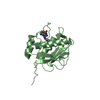
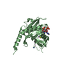
| Entry | Database: PDB / ID: 4es7 | ||||||
|---|---|---|---|---|---|---|---|
| Title | crystal structure of protein HC from Homo sapiens at 2 angstrom | ||||||
 Components Components | Protein AMBP | ||||||
 Keywords Keywords | IMMUNE SYSTEM | ||||||
| Function / homology |  Function and homology information Function and homology informationOxidoreductases; Acting on NADH or NADPH; With a heme protein as acceptor / calcium oxalate binding / IgA binding / negative regulation of immune response / heme catabolic process / negative regulation of JNK cascade / Scavenging of heme from plasma / calcium channel inhibitor activity / protein catabolic process / serine-type endopeptidase inhibitor activity ...Oxidoreductases; Acting on NADH or NADPH; With a heme protein as acceptor / calcium oxalate binding / IgA binding / negative regulation of immune response / heme catabolic process / negative regulation of JNK cascade / Scavenging of heme from plasma / calcium channel inhibitor activity / protein catabolic process / serine-type endopeptidase inhibitor activity / female pregnancy / : / carbohydrate binding / nuclear membrane / blood microparticle / oxidoreductase activity / cell adhesion / mitochondrial inner membrane / heme binding / cell surface / endoplasmic reticulum / protein homodimerization activity / extracellular space / extracellular exosome / extracellular region / plasma membrane / cytosol Similarity search - Function | ||||||
| Biological species |  Homo sapiens (human) Homo sapiens (human) | ||||||
| Method |  X-RAY DIFFRACTION / SYNCHROTRON / MOLECULAR REPLACEMENT / Resolution: 2.001 Å X-RAY DIFFRACTION / SYNCHROTRON / MOLECULAR REPLACEMENT / Resolution: 2.001 Å | ||||||
 Authors Authors | Zhang, Y.L. / Gao, Z.Q. / Wang, D.Q. / Dong, Y.H. | ||||||
 Citation Citation | Journal: To be Published Title: crystal structure of protein HC from Home sapiens at 2 angstrom Authors: Zhang, Y.L. / Gao, Z.Q. / Wang, D.Q. / Dong, Y.H. | ||||||
| History |
|
- Structure visualization
| Structure viewer | Molecule: MolmilJmol/JSmol |
|---|
- Downloads & links
-Download
| PDBx/mmCIF format | 4es7.cif.gz | 83.8 KB | Display | PDBx/mmCIF format |
|---|---|---|---|---|
| PDB format | pdb4es7.ent.gz | 62.2 KB | Display | PDB format |
| PDBx/mmJSON format | 4es7.json.gz | Tree view | PDBx/mmJSON format | |
| Others |  Other downloads Other downloads |
-Validation report
| Summary document | 4es7_validation.pdf.gz | 442.3 KB | Display | wwPDB validaton report |
|---|---|---|---|---|
| Full document | 4es7_full_validation.pdf.gz | 444 KB | Display | |
| Data in XML | 4es7_validation.xml.gz | 10.3 KB | Display | |
| Data in CIF | 4es7_validation.cif.gz | 12.8 KB | Display | |
| Arichive directory | https://data.pdbj.org/pub/pdb/validation_reports/es/4es7ftp://data.pdbj.org/pub/pdb/validation_reports/es/4es7 | HTTPS FTP |
-Related structure data
| Similar structure data |
|---|


-Links
 PDBj
PDBj


- Assembly
| Deposited unit | 
| ||||||||
|---|---|---|---|---|---|---|---|---|---|
| 1 |
| ||||||||
| Unit cell |
|
-Components
| #1: Protein | Mass: 22633.561 Da / Num. of mol.: 1 / Fragment: UNP residues 27-193 Source method: isolated from a genetically manipulated source Source: (gene. exp.) Homo sapiens (human) / Gene: AMBP, HCP, ITIL / Plasmid: pET28a / Production host:  | ||||
|---|---|---|---|---|---|
| #2: Chemical |   Mass: 106.120 Da / Num. of mol.: 3 / Source method: obtained synthetically / Formula: C4H10O3 Mass: 106.120 Da / Num. of mol.: 3 / Source method: obtained synthetically / Formula: C4H10O3#3: Water | ChemComp-HOH / |  Mass: 18.015 Da / Num. of mol.: 54 / Source method: isolated from a natural source / Formula: H2O Mass: 18.015 Da / Num. of mol.: 54 / Source method: isolated from a natural source / Formula: H2OHas protein modification | Y | |
-Experimental details
-Experiment
| Experiment | Method: X-RAY DIFFRACTION / Number of used crystals: 1 |
|---|
- Sample preparation
| Crystal | Density Matthews: 1.95 Å3/Da / Density % sol: 36.3 % |
|---|---|
| Crystal grow | Temperature: 293 K / Method: vapor diffusion, sitting drop / pH: 6.9 Details: 0.2M ammonium citrate, 20% PEG 3350, 0.1M HEPES pH 6.9, 40% 1,1,1,3,3,3-Hexafluoro-z-propanol, VAPOR DIFFUSION, SITTING DROP, temperature 293K |
-Data collection
| Diffraction | Mean temperature: 100 K |
|---|---|
| Diffraction source | Source: SYNCHROTRON / Site: SSRF  / Beamline: BL17U / Wavelength: 0.9791 Å / Beamline: BL17U / Wavelength: 0.9791 Å |
| Detector | Type: ADSC QUANTUM 315r / Detector: CCD / Date: Mar 20, 2011 |
| Radiation | Monochromator: double crystal monochromator / Protocol: SINGLE WAVELENGTH / Monochromatic (M) / Laue (L): M / Scattering type: x-ray |
| Radiation wavelength | Wavelength: 0.9791 Å / Relative weight: 1 |
| Reflection | Resolution: 2→50 Å / Num. all: 9932 / Num. obs: 9932 / % possible obs: 100 % / Observed criterion σ(F): 0 / Observed criterion σ(I): 0 / Redundancy: 7.1 % / Biso Wilson estimate: 30.63 Å2 / Rmerge(I) obs: 0.076 / Net I/σ(I): 50.3 |
| Reflection shell | Resolution: 2→2.07 Å / Redundancy: 7.3 % / Rmerge(I) obs: 0.383 / Mean I/σ(I) obs: 8.57 / Num. unique all: 982 / % possible all: 100 |
- Processing
| Software |
| ||||||||||||||||||||||||||||||||||||||||
|---|---|---|---|---|---|---|---|---|---|---|---|---|---|---|---|---|---|---|---|---|---|---|---|---|---|---|---|---|---|---|---|---|---|---|---|---|---|---|---|---|---|
| Refinement | Method to determine structure: MOLECULAR REPLACEMENT / Resolution: 2.001→34.669 Å / Occupancy max: 1 / Occupancy min: 1 / SU ML: 0.12 / σ(F): 1.34 / Phase error: 28.3 / Stereochemistry target values: ML
| ||||||||||||||||||||||||||||||||||||||||
| Solvent computation | Shrinkage radii: 0.6 Å / VDW probe radii: 0.9 Å / Solvent model: FLAT BULK SOLVENT MODEL / Bsol: 50.087 Å2 / ksol: 0.379 e/Å3 | ||||||||||||||||||||||||||||||||||||||||
| Displacement parameters | Biso max: 93.44 Å2 / Biso mean: 40.3096 Å2 / Biso min: 15.56 Å2
| ||||||||||||||||||||||||||||||||||||||||
| Refinement step | Cycle: LAST / Resolution: 2.001→34.669 Å
| ||||||||||||||||||||||||||||||||||||||||
| Refine LS restraints |
| ||||||||||||||||||||||||||||||||||||||||
| LS refinement shell | Refine-ID: X-RAY DIFFRACTION / Total num. of bins used: 3
| ||||||||||||||||||||||||||||||||||||||||
| Refinement TLS params. | Method: refined / Origin x: 14.9191 Å / Origin y: -6.7183 Å / Origin z: -9.2317 Å
| ||||||||||||||||||||||||||||||||||||||||
| Refinement TLS group |
|
