 Movie
Movie Controller


+ Open data

- Basic information
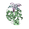
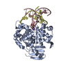
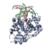
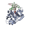
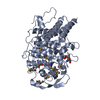
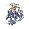
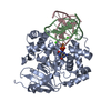
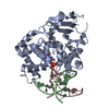
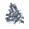
| Entry | Database: PDB / ID: 4ejz | ||||||
|---|---|---|---|---|---|---|---|
| Title | Structure of MBOgg1 in complex with low affinity DNA ligand | ||||||
 Components Components |
| ||||||
 Keywords Keywords | HYDROLASE/DNA / 8-oxoguanine DNA glycosylase / DNA / HYDROLASE-DNA complex | ||||||
| Function / homology |  Function and homology information Function and homology informationoxidized purine nucleobase lesion DNA N-glycosylase activity / class I DNA-(apurinic or apyrimidinic site) endonuclease activity / DNA-(apurinic or apyrimidinic site) lyase / nucleotide-excision repair / base-excision repair / damaged DNA binding Similarity search - Function | ||||||
| Biological species |  Thermoanaerobacter tengcongensis (bacteria) Thermoanaerobacter tengcongensis (bacteria) | ||||||
| Method |  X-RAY DIFFRACTION / SYNCHROTRON / MOLECULAR REPLACEMENT / Resolution: 3.05 Å X-RAY DIFFRACTION / SYNCHROTRON / MOLECULAR REPLACEMENT / Resolution: 3.05 Å | ||||||
 Authors Authors | Jiang, T. / Yu, H.J. / Bi, L.J. / Zhang, X.E. / Yang, M.Z. | ||||||
 Citation Citation | Journal: J.Struct.Biol. / Year: 2013 Title: Crystal structures of MBOgg1 in complex with two abasic DNA ligands Authors: Yu, H.J. / Yang, M.Z. / Zhang, X.E. / Bi, L.J. / Jiang, T. | ||||||
| History |
|
- Structure visualization
| Structure viewer | Molecule: MolmilJmol/JSmol |
|---|
- Downloads & links
-Download
| PDBx/mmCIF format | 4ejz.cif.gz | 159.2 KB | Display | PDBx/mmCIF format |
|---|---|---|---|---|
| PDB format | pdb4ejz.ent.gz | 121.1 KB | Display | PDB format |
| PDBx/mmJSON format | 4ejz.json.gz | Tree view | PDBx/mmJSON format | |
| Others |  Other downloads Other downloads |
-Validation report
| Arichive directory | https://data.pdbj.org/pub/pdb/validation_reports/ej/4ejzftp://data.pdbj.org/pub/pdb/validation_reports/ej/4ejz | HTTPS FTP |
|---|
-Related structure data
| Related structure data |  4ejySC S: Starting model for refinement C: citing same article ( |
|---|---|
| Similar structure data |

-Links
 PDBj
PDBj


- Assembly
| Deposited unit | 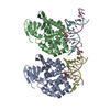
| ||||||||
|---|---|---|---|---|---|---|---|---|---|
| 1 | 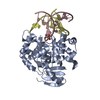
| ||||||||
| 2 | 
| ||||||||
| Unit cell |
|
-Components
| #1: Protein | Mass: 36189.273 Da / Num. of mol.: 2 Source method: isolated from a genetically manipulated source Source: (gene. exp.) Thermoanaerobacter tengcongensis (bacteria)Strain: MB4 / Gene: AlkA / Production host: References: UniProt: Q8R5T9, Hydrolases; Glycosylases; Hydrolysing N-glycosyl compounds #2: DNA chain | Mass: 4710.045 Da / Num. of mol.: 2 / Source method: obtained synthetically #3: DNA chain | Mass: 4954.214 Da / Num. of mol.: 2 / Source method: obtained synthetically |
|---|
-Experimental details
-Experiment
| Experiment | Method: X-RAY DIFFRACTION / Number of used crystals: 1 |
|---|
- Sample preparation
| Crystal | Density Matthews: 2.14 Å3/Da / Density % sol: 42.54 % |
|---|---|
| Crystal grow | Temperature: 289 K / Method: vapor diffusion, hanging drop Details: 20% PEG 3500, 0.1-0.15M KH2PO4, VAPOR DIFFUSION, HANGING DROP, temperature 289K |
-Data collection
| Diffraction | Mean temperature: 100 K |
|---|---|
| Diffraction source | Source: SYNCHROTRON / Site: SSRF  / Beamline: BL17U / Wavelength: 1.0089 Å / Beamline: BL17U / Wavelength: 1.0089 Å |
| Detector | Type: RAYONIX MX-225 / Detector: CCD / Date: Jan 18, 2011 |
| Radiation | Protocol: SINGLE WAVELENGTH / Monochromatic (M) / Laue (L): M / Scattering type: x-ray |
| Radiation wavelength | Wavelength: 1.0089 Å / Relative weight: 1 |
| Reflection | Resolution: 3.05→30 Å / Num. all: 14731 / Num. obs: 14731 / % possible obs: 99 % / Observed criterion σ(F): 0 / Observed criterion σ(I): 0 / Redundancy: 7 % / Rmerge(I) obs: 0.125 |
| Reflection shell | Resolution: 3.05→3.16 Å / Redundancy: 5.4 % / Rmerge(I) obs: 0.375 / Num. unique all: 1406 / % possible all: 93.8 |
- Processing
| Software |
| |||||||||||||||||||||||||||||||||||||||||||||||||||||||||||||||||
|---|---|---|---|---|---|---|---|---|---|---|---|---|---|---|---|---|---|---|---|---|---|---|---|---|---|---|---|---|---|---|---|---|---|---|---|---|---|---|---|---|---|---|---|---|---|---|---|---|---|---|---|---|---|---|---|---|---|---|---|---|---|---|---|---|---|---|
| Refinement | Method to determine structure: MOLECULAR REPLACEMENT Starting model: 4EJY Resolution: 3.05→30 Å / Cor.coef. Fo:Fc: 0.904 / Cor.coef. Fo:Fc free: 0.85 / Occupancy max: 1 / Occupancy min: 1 / Cross valid method: THROUGHOUT / ESU R Free: 0.596 / Stereochemistry target values: MAXIMUM LIKELIHOOD Details: HYDROGENS HAVE BEEN ADDED IN THE RIDING POSITIONS U VALUES: REFINED INDIVIDUALLY. THE STRUCTURE WAS REFINED ALSO WITH PHENIX.REFINE.
| |||||||||||||||||||||||||||||||||||||||||||||||||||||||||||||||||
| Solvent computation | Ion probe radii: 0.8 Å / Shrinkage radii: 0.8 Å / VDW probe radii: 1.4 Å / Solvent model: MASK | |||||||||||||||||||||||||||||||||||||||||||||||||||||||||||||||||
| Displacement parameters | Biso max: 100.23 Å2 / Biso mean: 51.488 Å2 / Biso min: 26.9 Å2
| |||||||||||||||||||||||||||||||||||||||||||||||||||||||||||||||||
| Refinement step | Cycle: LAST / Resolution: 3.05→30 Å
| |||||||||||||||||||||||||||||||||||||||||||||||||||||||||||||||||
| Refine LS restraints |
| |||||||||||||||||||||||||||||||||||||||||||||||||||||||||||||||||
| LS refinement shell | Resolution: 3.045→3.123 Å / Total num. of bins used: 20
|
