 Movie
Movie Controller

[English] 日本語
 Yorodumi
- PDB-4ee7: Crystal Structure of the Novel Phenazine Prenyltransferase EpzP i... -
+ Open data

- Basic information
| Entry | Database: PDB / ID: 4ee7 | ||||||
|---|---|---|---|---|---|---|---|
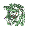
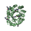
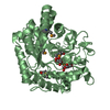
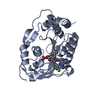
| Title | Crystal Structure of the Novel Phenazine Prenyltransferase EpzP in complex with S-thiolodiphosphate (methylated) | ||||||
 Components Components | Prenyltransferase | ||||||
 Keywords Keywords | TRANSFERASE / PT fold / dihydrophenazine carboxylate prenyltransferase | ||||||
| Function / homology | Aromatic prenyltransferase, CloQ-type / Prenyltransferase-like superfamily / Aromatic prenyltransferase Orf2 / Aromatic prenyltransferase / prenyltransferase activity / TRIHYDROGEN THIODIPHOSPHATE / Prenyltransferase Function and homology information Function and homology information | ||||||
| Biological species |  Streptomyces cinnamonensis (bacteria) Streptomyces cinnamonensis (bacteria) | ||||||
| Method |  X-RAY DIFFRACTION / SYNCHROTRON / PHASER / Resolution: 1.67 Å X-RAY DIFFRACTION / SYNCHROTRON / PHASER / Resolution: 1.67 Å | ||||||
 Authors Authors | Zocher, G. / Stehle, T. | ||||||
 Citation Citation | Journal: Plos One / Year: 2012 Title: Structure-based engineering increased the catalytic turnover rate of a novel phenazine prenyltransferase. Authors: Zocher, G. / Saleh, O. / Heim, J.B. / Herbst, D.A. / Heide, L. / Stehle, T. | ||||||
| History |
|
- Structure visualization
| Structure viewer | Molecule: MolmilJmol/JSmol |
|---|
- Downloads & links
-Download
| PDBx/mmCIF format | 4ee7.cif.gz | 248.9 KB | Display | PDBx/mmCIF format |
|---|---|---|---|---|
| PDB format | pdb4ee7.ent.gz | 200.7 KB | Display | PDB format |
| PDBx/mmJSON format | 4ee7.json.gz | Tree view | PDBx/mmJSON format | |
| Others |  Other downloads Other downloads |
-Validation report
| Arichive directory | https://data.pdbj.org/pub/pdb/validation_reports/ee/4ee7ftp://data.pdbj.org/pub/pdb/validation_reports/ee/4ee7 | HTTPS FTP |
|---|
-Related structure data


-Links
 PDBj
PDBj- Assembly
| Deposited unit | 
| ||||||||
|---|---|---|---|---|---|---|---|---|---|
| 1 | 
| ||||||||
| 2 | 
| ||||||||
| Unit cell |
|
-Components
-Protein , 1 types, 2 molecules AB
| #1: Protein | Mass: 33549.102 Da / Num. of mol.: 2 Source method: isolated from a genetically manipulated source Source: (gene. exp.) Streptomyces cinnamonensis (bacteria) / Gene: epzP / Production host: |
|---|
-Non-polymers , 5 types, 646 molecules 


| #2: Chemical | ChemComp-PIS /  Mass: 193.033 Da / Num. of mol.: 1 / Source method: obtained synthetically / Formula: H3O6P2S Mass: 193.033 Da / Num. of mol.: 1 / Source method: obtained synthetically / Formula: H3O6P2S | ||||||
|---|---|---|---|---|---|---|---|
| #3: Chemical | ChemComp-PG4 /  Mass: 194.226 Da / Num. of mol.: 4 / Source method: obtained synthetically / Formula: C8H18O5 / Comment: precipitant*YM Mass: 194.226 Da / Num. of mol.: 4 / Source method: obtained synthetically / Formula: C8H18O5 / Comment: precipitant*YM#4: Chemical | ChemComp-SO4 /  Mass: 96.063 Da / Num. of mol.: 6 / Source method: obtained synthetically / Formula: SO4 Mass: 96.063 Da / Num. of mol.: 6 / Source method: obtained synthetically / Formula: SO4#5: Chemical | ChemComp-CL / |  Mass: 35.453 Da / Num. of mol.: 1 / Source method: obtained synthetically / Formula: Cl Mass: 35.453 Da / Num. of mol.: 1 / Source method: obtained synthetically / Formula: Cl#6: Water | ChemComp-HOH / | Mass: 18.015 Da / Num. of mol.: 634 / Source method: isolated from a natural source / Formula: H2O |
-Details
| Has protein modification | Y |
|---|
-Experimental details
-Experiment
| Experiment | Method: X-RAY DIFFRACTION / Number of used crystals: 1 |
|---|
- Sample preparation
| Crystal | Density Matthews: 2.07 Å3/Da / Density % sol: 40.52 % |
|---|---|
| Crystal grow | Temperature: 293 K / Method: vapor diffusion, sitting drop / pH: 4.6 Details: 200 mM (NH4)2SO4, 30% (w/v) PEG2000MME, 100 mM sodium acetate 4.6, VAPOR DIFFUSION, SITTING DROP, temperature 293K |
-Data collection
| Diffraction | Mean temperature: 100 K |
|---|---|
| Diffraction source | Source: SYNCHROTRON / Site: SLS  / Beamline: X06DA / Wavelength: 1 Å / Beamline: X06DA / Wavelength: 1 Å |
| Detector | Type: MARMOSAIC 225 mm CCD / Detector: CCD / Date: May 7, 2010 |
| Radiation | Monochromator: DCCM / Protocol: SINGLE WAVELENGTH / Monochromatic (M) / Laue (L): M / Scattering type: x-ray |
| Radiation wavelength | Wavelength: 1 Å / Relative weight: 1 |
| Reflection | Resolution: 1.67→30 Å / Num. all: 65578 / Num. obs: 65469 / % possible obs: 99.8 % / Observed criterion σ(F): -3 / Observed criterion σ(I): -3 |
| Reflection shell | Resolution: 1.67→1.71 Å / % possible all: 99.4 |
- Processing
| Software |
| |||||||||||||||||||||||||||||||||||||||||||||||||||||||||||||||||||||||||||||||||||||
|---|---|---|---|---|---|---|---|---|---|---|---|---|---|---|---|---|---|---|---|---|---|---|---|---|---|---|---|---|---|---|---|---|---|---|---|---|---|---|---|---|---|---|---|---|---|---|---|---|---|---|---|---|---|---|---|---|---|---|---|---|---|---|---|---|---|---|---|---|---|---|---|---|---|---|---|---|---|---|---|---|---|---|---|---|---|---|
| Refinement | Method to determine structure: PHASER / Resolution: 1.67→29.39 Å / Cor.coef. Fo:Fc: 0.957 / Cor.coef. Fo:Fc free: 0.947 / SU B: 4.287 / SU ML: 0.076 / Cross valid method: THROUGHOUT / ESU R: 0.112 / ESU R Free: 0.109 / Stereochemistry target values: MAXIMUM LIKELIHOOD / Details: HYDROGENS HAVE BEEN ADDED IN THE RIDING POSITIONS
| |||||||||||||||||||||||||||||||||||||||||||||||||||||||||||||||||||||||||||||||||||||
| Solvent computation | Ion probe radii: 0.8 Å / Shrinkage radii: 0.8 Å / VDW probe radii: 1.4 Å / Solvent model: BABINET MODEL WITH MASK | |||||||||||||||||||||||||||||||||||||||||||||||||||||||||||||||||||||||||||||||||||||
| Displacement parameters | Biso mean: 15.188 Å2
| |||||||||||||||||||||||||||||||||||||||||||||||||||||||||||||||||||||||||||||||||||||
| Refinement step | Cycle: LAST / Resolution: 1.67→29.39 Å
| |||||||||||||||||||||||||||||||||||||||||||||||||||||||||||||||||||||||||||||||||||||
| Refine LS restraints |
| |||||||||||||||||||||||||||||||||||||||||||||||||||||||||||||||||||||||||||||||||||||
| LS refinement shell | Resolution: 1.67→1.713 Å / Total num. of bins used: 20
| |||||||||||||||||||||||||||||||||||||||||||||||||||||||||||||||||||||||||||||||||||||
| Refinement TLS params. | Method: refined / Refine-ID: X-RAY DIFFRACTION
| |||||||||||||||||||||||||||||||||||||||||||||||||||||||||||||||||||||||||||||||||||||
| Refinement TLS group |
|
