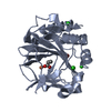
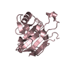
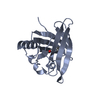
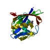
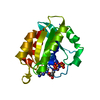
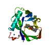
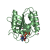
- PDB-4dzr: The crystal structure of protein-(glutamine-N5) methyltransferase... -
+
Open data
ID or keywords:
Loading...
-
Basic information
Entry
Database: PDB / ID: 4dzr
Title
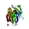
The crystal structure of protein-(glutamine-N5) methyltransferase (release factor-specific) from Alicyclobacillus acidocaldarius subsp. acidocaldarius DSM 446
Mass: 18.015 Da / Num. of mol.: 5 / Source method: isolated from a natural source / Formula: H2O
Has protein modification
Y
Sequence details
THE AUTHORS STATE THAT THE EXPRESSED PROTEIN WAS A FULL LENGTH PROTEIN WITH SEQUENCE: SNA(MSE) ...THE AUTHORS STATE THAT THE EXPRESSED PROTEIN WAS A FULL LENGTH PROTEIN WITH SEQUENCE: SNA(MSE)SEAKYFVARLLKAIAEQLPQSPAYRALPLDE RKRLAEREAEQIVAHALGWDRVKLLQSLGDEVPDEIAERAARLAALRVQGEPLAYVLGKQDF YGRTFEVGPDCLIPRPDTEVLVEEAIRFLKR(MSE)PSGTRVIDVGTGSGCIAVSIALACPG VSVTAVDLS(MSE)DALAVARRNAERFGAVVDWAAADGIEWLIERAERGRPWHAIVSNPPYI PTGEIDQLEPSVRDYEPRLALDGGEDGLQFYRR(MSE)AALPPYVLARGRAGVFLEVGHNQA DEVARLFAPWRERGFRVRKVKDLRGIDRVIAVTREPGSPPESENL AND SINCE TRYPSIN WAS ADDED IN PROTEIN FOR CRYSTALLIZATION PURPOSES, THE PROTEIN WAS CHOPPED AT MULTIPLE SITES. ESPECIALLY, AN EXPECTED N-TERMINAL DOMAIN WAS COMPLETELY GONE AND SEVERAL LOOPS ARE MISSING IN THE STRUCTURE. HOWEVER, THE AUTHORS DO NOT KNOW EXACTLY WHERE THE SEQUENCE STARTS.
-
Experimental details
-
Experiment
Experiment
Method: X-RAY DIFFRACTION / Number of used crystals: 1
-
Sample preparation
Crystal
Density Matthews: 2.45 Å3/Da / Density % sol: 49.85 %
In the structure databanks used in Yorodumi, some data are registered as the other names, "COVID-19 virus" and "2019-nCoV". Here are the details of the virus and the list of structure data.
Jan 31, 2019. EMDB accession codes are about to change! (news from PDBe EMDB page)
EMDB accession codes are about to change! (news from PDBe EMDB page)
The allocation of 4 digits for EMDB accession codes will soon come to an end. Whilst these codes will remain in use, new EMDB accession codes will include an additional digit and will expand incrementally as the available range of codes is exhausted. The current 4-digit format prefixed with “EMD-” (i.e. EMD-XXXX) will advance to a 5-digit format (i.e. EMD-XXXXX), and so on. It is currently estimated that the 4-digit codes will be depleted around Spring 2019, at which point the 5-digit format will come into force.
The EM Navigator/Yorodumi systems omit the EMD- prefix.
Related info.:Q: What is EMD? / ID/Accession-code notation in Yorodumi/EM Navigator
Yorodumi is a browser for structure data from EMDB, PDB, SASBDB, etc.
This page is also the successor to EM Navigator detail page, and also detail information page/front-end page for Omokage search.
The word "yorodu" (or yorozu) is an old Japanese word meaning "ten thousand". "mi" (miru) is to see.
Related info.:EMDB / PDB / SASBDB / Comparison of 3 databanks / Yorodumi Search / Aug 31, 2016. New EM Navigator & Yorodumi / Yorodumi Papers / Jmol/JSmol / Function and homology information / Changes in new EM Navigator and Yorodumi
 Movie
Movie Controller

 Yorodumi
 Open data
 Basic information
 Components
 Keywords
 Function and homology information
 Alicyclobacillus acidocaldarius subsp. acidocaldarius DSM 446 (bacteria)
 X-RAY DIFFRACTION /
  Authors
 Citation
 Structure visualization
 Downloads & links
 Other downloads
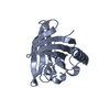

 PDBj
PDBj Assembly

 Mass: 40.078 Da / Num. of mol.: 1 / Source method: obtained synthetically / Formula: Ca
 Mass: 59.044 Da / Num. of mol.: 1 / Source method: obtained synthetically / Formula: C2H3O2
 Mass: 92.094 Da / Num. of mol.: 1 / Source method: obtained synthetically / Formula: C3H8O3
 Mass: 18.015 Da / Num. of mol.: 5 / Source method: isolated from a natural source / Formula: H2O
 Sample preparation
 / Beamline: 19-ID / Wavelength: 0.97931, 0.97948
 Processing