 Movie
Movie Controller

[English] 日本語
 Yorodumi
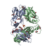
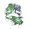
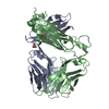
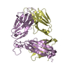
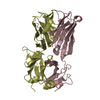
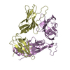
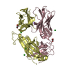
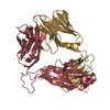
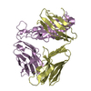
- PDB-4dzb: Mucosal-associated invariant T cell receptor, Valpha7.2Jalpha33-Vbeta2 -
+ Open data

- Basic information
| Entry | Database: PDB / ID: 4dzb | ||||||
|---|---|---|---|---|---|---|---|
| Title | Mucosal-associated invariant T cell receptor, Valpha7.2Jalpha33-Vbeta2 | ||||||
 Components Components |
| ||||||
 Keywords Keywords | IMMUNE SYSTEM / MAIT T cell receptor | ||||||
| Function / homology | Immunoglobulins / Immunoglobulin-like / Sandwich / Mainly Beta Function and homology information Function and homology information | ||||||
| Biological species |  Homo sapiens (human) Homo sapiens (human) | ||||||
| Method |  X-RAY DIFFRACTION / SYNCHROTRON / MOLECULAR REPLACEMENT / Resolution: 1.7 Å X-RAY DIFFRACTION / SYNCHROTRON / MOLECULAR REPLACEMENT / Resolution: 1.7 Å | ||||||
 Authors Authors | Patel, O. / Rossjohn, J. | ||||||
 Citation Citation | Journal: J.Exp.Med. / Year: 2012 Title: Structural insight into MR1-mediated recognition of the mucosal associated invariant T cell receptor. Authors: Reantragoon, R. / Kjer-Nielsen, L. / Patel, O. / Chen, Z. / Illing, P.T. / Bhati, M. / Kostenko, L. / Bharadwaj, M. / Meehan, B. / Hansen, T.H. / Godfrey, D.I. / Rossjohn, J. / McCluskey, J. | ||||||
| History |
|
- Structure visualization
| Structure viewer | Molecule: MolmilJmol/JSmol |
|---|
- Downloads & links
-Download
| PDBx/mmCIF format | 4dzb.cif.gz | 103.5 KB | Display | PDBx/mmCIF format |
|---|---|---|---|---|
| PDB format | pdb4dzb.ent.gz | 79.6 KB | Display | PDB format |
| PDBx/mmJSON format | 4dzb.json.gz | Tree view | PDBx/mmJSON format | |
| Others |  Other downloads Other downloads |
-Validation report
| Arichive directory | https://data.pdbj.org/pub/pdb/validation_reports/dz/4dzbftp://data.pdbj.org/pub/pdb/validation_reports/dz/4dzb | HTTPS FTP |
|---|
-Related structure data
| Related structure data |  2nx5S S: Starting model for refinement |
|---|---|
| Similar structure data |

-Links
 PDBj
PDBj- Assembly
| Deposited unit | 
| ||||||||
|---|---|---|---|---|---|---|---|---|---|
| 1 |
| ||||||||
| Unit cell |
|
-Components
| #1: Protein | Mass: 22700.131 Da / Num. of mol.: 1 Source method: isolated from a genetically manipulated source Source: (gene. exp.) Homo sapiens (human) / Plasmid: pET30b / Production host:  |
|---|---|
| #2: Protein | Mass: 27622.797 Da / Num. of mol.: 1 Source method: isolated from a genetically manipulated source Source: (gene. exp.) Homo sapiens (human) / Plasmid: pET30b / Production host: |
| #3: Water | ChemComp-HOH /  Mass: 18.015 Da / Num. of mol.: 271 / Source method: isolated from a natural source / Formula: H2O Mass: 18.015 Da / Num. of mol.: 271 / Source method: isolated from a natural source / Formula: H2O |
| Has protein modification | Y |
-Experimental details
-Experiment
| Experiment | Method: X-RAY DIFFRACTION / Number of used crystals: 1 |
|---|
- Sample preparation
| Crystal | Density Matthews: 2.09 Å3/Da / Density % sol: 41.04 % |
|---|---|
| Crystal grow | Temperature: 298 K / Method: vapor diffusion, hanging drop / pH: 5 Details: 25% PEG 1500, 0.1M MIB buffer (sodium malonate, imidazole, boric acid), VAPOR DIFFUSION, HANGING DROP, temperature 298K |
-Data collection
| Diffraction | Mean temperature: 100 K |
|---|---|
| Diffraction source | Source: SYNCHROTRON / Site: Australian Synchrotron  / Beamline: MX2 / Wavelength: 0.95453 Å / Beamline: MX2 / Wavelength: 0.95453 Å |
| Detector | Type: ADSC QUANTUM 315r / Detector: CCD / Date: Nov 10, 2005 |
| Radiation | Protocol: SINGLE WAVELENGTH / Monochromatic (M) / Laue (L): M / Scattering type: x-ray |
| Radiation wavelength | Wavelength: 0.95453 Å / Relative weight: 1 |
| Reflection | Resolution: 1.7→50 Å / Num. all: 45957 / Num. obs: 44624 / % possible obs: 97.1 % / Redundancy: 7.6 % / Biso Wilson estimate: 17.5 Å2 / Rmerge(I) obs: 0.093 / Net I/σ(I): 13.7 |
| Reflection shell | Resolution: 1.7→1.79 Å / Redundancy: 7.4 % / Rmerge(I) obs: 0.773 / Mean I/σ(I) obs: 2.5 / Num. unique all: 6281 / % possible all: 92.6 |
- Processing
| Software |
| |||||||||||||||||||||||||||||||||||||||||||||||||||||||||||||||||
|---|---|---|---|---|---|---|---|---|---|---|---|---|---|---|---|---|---|---|---|---|---|---|---|---|---|---|---|---|---|---|---|---|---|---|---|---|---|---|---|---|---|---|---|---|---|---|---|---|---|---|---|---|---|---|---|---|---|---|---|---|---|---|---|---|---|---|
| Refinement | Method to determine structure: MOLECULAR REPLACEMENT Starting model: pdb entry 2NX5 Resolution: 1.7→50 Å / Cor.coef. Fo:Fc: 0.954 / Cor.coef. Fo:Fc free: 0.93 / SU B: 2.115 / SU ML: 0.071 / Cross valid method: THROUGHOUT / σ(F): 0 / σ(I): 0 / ESU R: 0.121 / ESU R Free: 0.118 / Stereochemistry target values: MAXIMUM LIKELIHOOD / Details: HYDROGENS HAVE BEEN ADDED IN THE RIDING POSITIONS
| |||||||||||||||||||||||||||||||||||||||||||||||||||||||||||||||||
| Solvent computation | Ion probe radii: 0.8 Å / Shrinkage radii: 0.8 Å / VDW probe radii: 1.4 Å / Solvent model: MASK | |||||||||||||||||||||||||||||||||||||||||||||||||||||||||||||||||
| Displacement parameters | Biso mean: 18.68 Å2
| |||||||||||||||||||||||||||||||||||||||||||||||||||||||||||||||||
| Refinement step | Cycle: LAST / Resolution: 1.7→50 Å
| |||||||||||||||||||||||||||||||||||||||||||||||||||||||||||||||||
| Refine LS restraints |
| |||||||||||||||||||||||||||||||||||||||||||||||||||||||||||||||||
| LS refinement shell | Resolution: 1.7→1.744 Å / Total num. of bins used: 20
|
