 Movie
Movie Controller

[English] 日本語
 Yorodumi
- PDB-4did: Crystal structure of Salmonella effector N-terminal domain SopB i... -
+ Open data

- Basic information
| Entry | Database: PDB / ID: 4did | ||||||
|---|---|---|---|---|---|---|---|
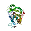
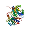
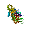
| Title | Crystal structure of Salmonella effector N-terminal domain SopB in complex with Cdc42 | ||||||
 Components Components |
| ||||||
 Keywords Keywords | HYDROLASE/HYDROLASE / small GTPase / GTP binding / HYDROLASE-HYDROLASE complex | ||||||
| Function / homology |  Function and homology information Function and homology informationsymbiont-mediated perturbation of host vacuole organization / symbiont-mediated suppression of host reactive oxygen species generation / symbiont-containing vacuole / negative regulation of signaling / GBD domain binding / positive regulation of pinocytosis / COG complex / endothelin receptor signaling pathway involved in heart process / regulation of protein kinase activity / cardiac neural crest cell migration involved in outflow tract morphogenesis ...symbiont-mediated perturbation of host vacuole organization / symbiont-mediated suppression of host reactive oxygen species generation / symbiont-containing vacuole / negative regulation of signaling / GBD domain binding / positive regulation of pinocytosis / COG complex / endothelin receptor signaling pathway involved in heart process / regulation of protein kinase activity / cardiac neural crest cell migration involved in outflow tract morphogenesis / storage vacuole / dendritic cell migration / neuron fate determination / apolipoprotein A-I receptor binding / positive regulation of epithelial cell proliferation involved in lung morphogenesis / regulation of attachment of spindle microtubules to kinetochore / organelle transport along microtubule / lipid phosphatase activity / Inactivation of CDC42 and RAC1 / positive regulation of pseudopodium assembly / host-mediated perturbation of viral process / regulation of filopodium assembly / cardiac conduction system development / leading edge membrane / neuropilin signaling pathway / Hydrolases; Acting on ester bonds; Phosphoric-monoester hydrolases / establishment of Golgi localization / filopodium assembly / cell junction assembly / establishment of epithelial cell apical/basal polarity / dendritic spine morphogenesis / adherens junction organization / GTP-dependent protein binding / thioesterase binding / regulation of lamellipodium assembly / regulation of stress fiber assembly / embryonic heart tube development / RHO GTPases activate KTN1 / DCC mediated attractive signaling / CD28 dependent Vav1 pathway / regulation of postsynapse organization / positive regulation of filopodium assembly / Wnt signaling pathway, planar cell polarity pathway / phagocytosis, engulfment / RHOV GTPase cycle / regulation of mitotic nuclear division / Myogenesis / nuclear migration / small GTPase-mediated signal transduction / symbiont-mediated suppression of host TRAF-mediated signal transduction / positive regulation of cytokinesis / spindle midzone / RHOJ GTPase cycle / heart contraction / RHOQ GTPase cycle / establishment of cell polarity / Golgi organization / RHOU GTPase cycle / establishment or maintenance of cell polarity / RHO GTPases activate PAKs / CDC42 GTPase cycle / macrophage differentiation / RHOG GTPase cycle / RAC3 GTPase cycle / RAC2 GTPase cycle / RHO GTPases Activate WASPs and WAVEs / negative regulation of protein-containing complex assembly / RHO GTPases activate IQGAPs / GPVI-mediated activation cascade / positive regulation of lamellipodium assembly / phagocytic vesicle / positive regulation of stress fiber assembly / RAC1 GTPase cycle / EPHB-mediated forward signaling / positive regulation of substrate adhesion-dependent cell spreading / substantia nigra development / Gene and protein expression by JAK-STAT signaling after Interleukin-12 stimulation / positive regulation of DNA replication / actin filament organization / integrin-mediated signaling pathway / small monomeric GTPase / cell periphery / regulation of actin cytoskeleton organization / FCGR3A-mediated phagocytosis / filopodium / EGFR downregulation / RHO GTPases Activate Formins / negative regulation of cell growth / Regulation of actin dynamics for phagocytic cup formation / MAPK6/MAPK4 signaling / VEGFA-VEGFR2 Pathway / cellular response to type II interferon / endocytosis / apical part of cell / cytoplasmic ribonucleoprotein granule / cell-cell junction / G beta:gamma signalling through CDC42 / mitotic spindle / intracellular protein localization / ubiquitin protein ligase activity Similarity search - Function | ||||||
| Biological species |  Homo sapiens (human) Homo sapiens (human) Salmonella enterica subsp. enterica serovar Typhimurium (bacteria) Salmonella enterica subsp. enterica serovar Typhimurium (bacteria) | ||||||
| Method |  X-RAY DIFFRACTION / SYNCHROTRON / MOLECULAR REPLACEMENT / Resolution: 2.3501 Å X-RAY DIFFRACTION / SYNCHROTRON / MOLECULAR REPLACEMENT / Resolution: 2.3501 Å | ||||||
 Authors Authors | Burkinshaw, B.J. / Strynadka, N.C.J. | ||||||
 Citation Citation | Journal: J.Biol.Chem. / Year: 2012 Title: Structure of Salmonella Effector Protein SopB N-terminal Domain in Complex with Host Rho GTPase Cdc42. Authors: Burkinshaw, B.J. / Prehna, G. / Worrall, L.J. / Strynadka, N.C. | ||||||
| History |
|
- Structure visualization
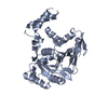
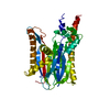
| Structure viewer | Molecule: MolmilJmol/JSmol |
|---|
- Downloads & links
-Download
| PDBx/mmCIF format | 4did.cif.gz | 77.6 KB | Display | PDBx/mmCIF format |
|---|---|---|---|---|
| PDB format | pdb4did.ent.gz | 56 KB | Display | PDB format |
| PDBx/mmJSON format | 4did.json.gz | Tree view | PDBx/mmJSON format | |
| Others |  Other downloads Other downloads |
-Validation report
| Arichive directory | https://data.pdbj.org/pub/pdb/validation_reports/di/4didftp://data.pdbj.org/pub/pdb/validation_reports/di/4did | HTTPS FTP |
|---|
-Related structure data
| Related structure data | 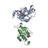 1an0S S: Starting model for refinement |
|---|---|
| Similar structure data |


-Links
 PDBj
PDBj


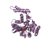
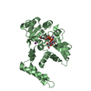
- Assembly
| Deposited unit | 
| ||||||||
|---|---|---|---|---|---|---|---|---|---|
| 1 |
| ||||||||
| Unit cell |
|
-Components
| #1: Protein | Mass: 21515.635 Da / Num. of mol.: 1 Source method: isolated from a genetically manipulated source Source: (gene. exp.) Homo sapiens (human) / Gene: CDC42 / Plasmid: pET28 / Production host: |
|---|---|
| #2: Protein | Mass: 16781.119 Da / Num. of mol.: 1 / Fragment: UNP residues 30-181 Source method: isolated from a genetically manipulated source Source: (gene. exp.) Salmonella enterica subsp. enterica serovar Typhimurium (bacteria)Gene: sigD, sopB, STM1091 / Plasmid: pET21 / Production host: References: UniProt: O30916, Hydrolases; Acting on ester bonds; Phosphoric-monoester hydrolases |
| #3: Chemical | ChemComp-GDP /   Type: RNA linking / Mass: 443.201 Da / Num. of mol.: 1 / Source method: obtained synthetically / Formula: C10H15N5O11P2 / Comment: GDP, energy-carrying molecule*YM Type: RNA linking / Mass: 443.201 Da / Num. of mol.: 1 / Source method: obtained synthetically / Formula: C10H15N5O11P2 / Comment: GDP, energy-carrying molecule*YM |
| #4: Chemical | ChemComp-MG /   Mass: 24.305 Da / Num. of mol.: 1 / Source method: obtained synthetically / Formula: Mg Mass: 24.305 Da / Num. of mol.: 1 / Source method: obtained synthetically / Formula: Mg |
| #5: Water | ChemComp-HOH /  Mass: 18.015 Da / Num. of mol.: 71 / Source method: isolated from a natural source / Formula: H2O Mass: 18.015 Da / Num. of mol.: 71 / Source method: isolated from a natural source / Formula: H2O |
-Experimental details
-Experiment
| Experiment | Method: X-RAY DIFFRACTION / Number of used crystals: 1 |
|---|
- Sample preparation
| Crystal | Density Matthews: 3.27 Å3/Da / Density % sol: 62.33 % |
|---|---|
| Crystal grow | Temperature: 277 K / Method: vapor diffusion, sitting drop / pH: 4.2 Details: 0.2M sodium chloride, 0.1M phosphate-citrate pH 4.2, 20% PEG 8000, VAPOR DIFFUSION, SITTING DROP, temperature 277K |
-Data collection
| Diffraction | Mean temperature: 200 K |
|---|---|
| Diffraction source | Source: SYNCHROTRON / Site: CLSI  / Beamline: 08B1-1 / Wavelength: 0.9795 Å / Beamline: 08B1-1 / Wavelength: 0.9795 Å |
| Detector | Type: RAYONIX MX300HE / Detector: CCD / Date: Jul 29, 2010 |
| Radiation | Monochromator: Si 111 / Protocol: SINGLE WAVELENGTH / Monochromatic (M) / Laue (L): M / Scattering type: x-ray |
| Radiation wavelength | Wavelength: 0.9795 Å / Relative weight: 1 |
| Reflection | Resolution: 2.35→47.8 Å / Num. all: 21708 / Num. obs: 21675 / % possible obs: 99.7 % / Observed criterion σ(F): 5.8 / Observed criterion σ(I): 6.2 |
| Reflection shell | Resolution: 2.35→2.45 Å / % possible all: 100 |
- Processing
| Software |
| |||||||||||||||||||||||||||||||||||||||||||||||||||||||||||||||
|---|---|---|---|---|---|---|---|---|---|---|---|---|---|---|---|---|---|---|---|---|---|---|---|---|---|---|---|---|---|---|---|---|---|---|---|---|---|---|---|---|---|---|---|---|---|---|---|---|---|---|---|---|---|---|---|---|---|---|---|---|---|---|---|---|
| Refinement | Method to determine structure: MOLECULAR REPLACEMENT Starting model: PDB ENTRY 1AN0 Resolution: 2.3501→45.617 Å / SU ML: 0.3 / σ(F): 1.34 / Phase error: 25.46 / Stereochemistry target values: ML
| |||||||||||||||||||||||||||||||||||||||||||||||||||||||||||||||
| Solvent computation | Shrinkage radii: 0.86 Å / VDW probe radii: 1.1 Å / Solvent model: FLAT BULK SOLVENT MODEL / Bsol: 44.346 Å2 / ksol: 0.354 e/Å3 | |||||||||||||||||||||||||||||||||||||||||||||||||||||||||||||||
| Displacement parameters |
| |||||||||||||||||||||||||||||||||||||||||||||||||||||||||||||||
| Refinement step | Cycle: LAST / Resolution: 2.3501→45.617 Å
| |||||||||||||||||||||||||||||||||||||||||||||||||||||||||||||||
| Refine LS restraints |
| |||||||||||||||||||||||||||||||||||||||||||||||||||||||||||||||
| LS refinement shell |
|
