| 登録情報 | データベース: PDB / ID: 4dc2
|
|---|
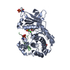
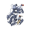
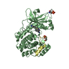
| タイトル | Structure of PKC in Complex with a Substrate Peptide from Par-3 |
|---|
 要素 要素 | - Partitioning defective 3 homolog
- Protein kinase C iota type
|
|---|
 キーワード キーワード | TRANSFERASE/TRANSFERASE SUBSTRATE / Kinase / substrate / Cell polarity / Par-3 / atypical PKC / TRANSFERASE-TRANSFERASE SUBSTRATE complex |
|---|
| 機能・相同性 |  機能・相同性情報 機能・相同性情報
Pre-NOTCH Transcription and Translation / Tight junction interactions / Tight junction interactions / regulation of actin filament-based process / TGF-beta receptor signaling in EMT (epithelial to mesenchymal transition) / internode region of axon / regulation of cellular localization / p75NTR recruits signalling complexes / Golgi vesicle budding / diacylglycerol-dependent, calcium-independent serine/threonine kinase activity ...Pre-NOTCH Transcription and Translation / Tight junction interactions / Tight junction interactions / regulation of actin filament-based process / TGF-beta receptor signaling in EMT (epithelial to mesenchymal transition) / internode region of axon / regulation of cellular localization / p75NTR recruits signalling complexes / Golgi vesicle budding / diacylglycerol-dependent, calcium-independent serine/threonine kinase activity / apical constriction / PAR polarity complex / establishment of centrosome localization / protein kinase C / establishment of epithelial cell polarity / establishment of apical/basal cell polarity / lateral loop / negative regulation of glial cell apoptotic process / eye photoreceptor cell development / bicellular tight junction assembly / positive regulation of myelination / Schmidt-Lanterman incisure / negative regulation of peptidyl-threonine phosphorylation / establishment or maintenance of epithelial cell apical/basal polarity / myelination in peripheral nervous system / KEAP1-NFE2L2 pathway / phosphatidylinositol-3-phosphate binding / cell-cell junction organization / wound healing, spreading of cells / protein targeting to membrane / centrosome localization / apical junction complex / establishment of cell polarity / cell leading edge / phosphatidylinositol-3,4,5-trisphosphate binding / brush border / positive regulation of receptor internalization / positive regulation of glial cell proliferation / positive regulation of endothelial cell apoptotic process / bicellular tight junction / axonal growth cone / regulation of postsynaptic membrane neurotransmitter receptor levels / intercellular bridge / phosphatidylinositol-4,5-bisphosphate binding / phosphatidylinositol binding / endomembrane system / response to interleukin-1 / actin filament organization / positive regulation of D-glucose import / positive regulation of protein localization to plasma membrane / adherens junction / positive regulation of NF-kappaB transcription factor activity / positive regulation of neuron projection development / phospholipid binding / microtubule cytoskeleton organization / spindle / Schaffer collateral - CA1 synapse / cellular response to insulin stimulus / apical part of cell / cell-cell junction / cell junction / intracellular protein localization / cell migration / microtubule cytoskeleton / cell cortex / protein phosphatase binding / negative regulation of neuron apoptotic process / protein kinase activity / cell adhesion / endosome / cilium / apical plasma membrane / Golgi membrane / cell division / protein serine kinase activity / neuronal cell body / protein serine/threonine kinase activity / glutamatergic synapse / protein-containing complex / zinc ion binding / ATP binding / identical protein binding / nucleus / cytoplasm / cytosol類似検索 - 分子機能 Par3/HAL, N-terminal / N-terminal of Par3 and HAL proteins / : / Atypical protein kinase C iota type, catalytic domain / Protein kinase C / Protein kinase C, PB1 domain / PB1 domain / PB1 domain / PB1 domain / : ...Par3/HAL, N-terminal / N-terminal of Par3 and HAL proteins / : / Atypical protein kinase C iota type, catalytic domain / Protein kinase C / Protein kinase C, PB1 domain / PB1 domain / PB1 domain / PB1 domain / : / PB1 domain profile. / Protein kinase, C-terminal / Protein kinase C terminal domain / Diacylglycerol/phorbol-ester binding / Phorbol esters/diacylglycerol binding domain (C1 domain) / Zinc finger phorbol-ester/DAG-type signature. / Zinc finger phorbol-ester/DAG-type profile. / Protein kinase C conserved region 1 (C1) domains (Cysteine-rich domains) / Protein kinase C-like, phorbol ester/diacylglycerol-binding domain / C1-like domain superfamily / Extension to Ser/Thr-type protein kinases / AGC-kinase, C-terminal / AGC-kinase C-terminal domain profile. / PDZ domain / PDZ domain profile. / Domain present in PSD-95, Dlg, and ZO-1/2. / PDZ domain / PDZ superfamily / Phosphorylase Kinase; domain 1 / Phosphorylase Kinase; domain 1 / Transferase(Phosphotransferase) domain 1 / Transferase(Phosphotransferase); domain 1 / Serine/threonine-protein kinase, active site / Serine/Threonine protein kinases active-site signature. / Protein kinase domain / Serine/Threonine protein kinases, catalytic domain / Protein kinase, ATP binding site / Protein kinases ATP-binding region signature. / Protein kinase domain profile. / Protein kinase domain / Protein kinase-like domain superfamily / 2-Layer Sandwich / Orthogonal Bundle / Mainly Alpha / Alpha Beta類似検索 - ドメイン・相同性 ADENINE / Protein kinase C iota type / Partitioning defective 3 homolog類似検索 - 構成要素 |
|---|
| 生物種 |   Mus musculus (ハツカネズミ) Mus musculus (ハツカネズミ)
 Rattus norvegicus (ドブネズミ) Rattus norvegicus (ドブネズミ) |
|---|
| 手法 |  X線回折 / シンクロトロン / 分子置換 / 解像度: 2.4 Å X線回折 / シンクロトロン / 分子置換 / 解像度: 2.4 Å |
|---|
 データ登録者 データ登録者 | Shang, Y. / Wang, C. / Yu, J. / Zhang, M. |
|---|
 引用 引用 | ジャーナル: Structure / 年: 2012
タイトル: Substrate recognition mechanism of atypical protein kinase Cs revealed by the structure of PKC iota in complex with a substrate peptide from Par-3
著者: Wang, C. / Shang, Y. / Yu, J. / Zhang, M. |
|---|
| 履歴 | | 登録 | 2012年1月17日 | 登録サイト: RCSB / 処理サイト: PDBJ |
|---|
| 改定 1.0 | 2012年7月11日 | Provider: repository / タイプ: Initial release |
|---|
| 改定 1.1 | 2024年11月20日 | Group: Data collection / Database references ...Data collection / Database references / Derived calculations / Structure summary
カテゴリ: chem_comp_atom / chem_comp_bond ...chem_comp_atom / chem_comp_bond / database_2 / pdbx_entry_details / pdbx_modification_feature / struct_conn / struct_ref_seq_dif / struct_site
Item: _database_2.pdbx_DOI / _database_2.pdbx_database_accession ..._database_2.pdbx_DOI / _database_2.pdbx_database_accession / _struct_conn.pdbx_leaving_atom_flag / _struct_ref_seq_dif.details / _struct_site.pdbx_auth_asym_id / _struct_site.pdbx_auth_comp_id / _struct_site.pdbx_auth_seq_id |
|---|
|
|---|
 ムービー
ムービー コントローラー


 データを開く
 基本情報
基本情報 構造の表示
構造の表示 ダウンロードとリンク
 その他のダウンロード


 PDBj
PDBj


 集合体
集合体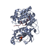

 Spodoptera frugiperda (ツマジロクサヨトウ)
 分子量: 135.127 Da / 分子数: 1 / 由来タイプ: 合成 / 式: C5H5N5
 分子量: 18.015 Da / 分子数: 52 / 由来タイプ: 天然 / 式: H2O
 試料調製
試料調製 / ビームライン: BL17U
 解析
解析