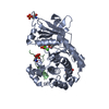
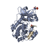
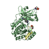
[English] 日本語
Yorodumi
- PDB-4dc2: Structure of PKC in Complex with a Substrate Peptide from Par-3 -

+
Open data


ID or keywords:

Loading...

-
Basic information

Entry
Database: PDB / ID: 4dc2
TitleStructure of PKC in Complex with a Substrate Peptide from Par-3
Components
  • Partitioning defective 3 homolog
  • Protein kinase C iota type
KeywordsTRANSFERASE/TRANSFERASE SUBSTRATE / Kinase / substrate / Cell polarity / Par-3 / atypical PKC / TRANSFERASE-TRANSFERASE SUBSTRATE complex
Function / homology
Function and homology information


Pre-NOTCH Transcription and Translation / Tight junction interactions / Tight junction interactions / regulation of actin filament-based process / TGF-beta receptor signaling in EMT (epithelial to mesenchymal transition) / internode region of axon / regulation of cellular localization / calcium,diacylglycerol-dependent serine/threonine kinase activity / Golgi vesicle budding / PAR polarity complex ...Pre-NOTCH Transcription and Translation / Tight junction interactions / Tight junction interactions / regulation of actin filament-based process / TGF-beta receptor signaling in EMT (epithelial to mesenchymal transition) / internode region of axon / regulation of cellular localization / calcium,diacylglycerol-dependent serine/threonine kinase activity / Golgi vesicle budding / PAR polarity complex / protein localization => GO:0008104 / p75NTR recruits signalling complexes / apical constriction / establishment of apical/basal cell polarity / establishment of centrosome localization / protein kinase C / negative regulation of glial cell apoptotic process / diacylglycerol-dependent serine/threonine kinase activity / positive regulation of myelination / lateral loop / establishment of epithelial cell polarity / eye photoreceptor cell development / Schmidt-Lanterman incisure / myelination in peripheral nervous system / bicellular tight junction assembly / cell-cell junction organization / phosphatidylinositol-3-phosphate binding / establishment or maintenance of epithelial cell apical/basal polarity / positive regulation of endothelial cell apoptotic process / protein targeting to membrane / wound healing, spreading of cells / centrosome localization / apical junction complex / establishment of cell polarity / intercellular bridge / negative regulation of peptidyl-threonine phosphorylation / cell leading edge / regulation of postsynaptic membrane neurotransmitter receptor levels / phosphatidylinositol-3,4,5-trisphosphate binding / positive regulation of receptor internalization / bicellular tight junction / endomembrane system / axonal growth cone / positive regulation of glial cell proliferation / phosphatidylinositol-4,5-bisphosphate binding / phosphatidylinositol binding / response to interleukin-1 / actin filament organization / phospholipid binding / positive regulation of protein localization to plasma membrane / adherens junction / positive regulation of glucose import / Schaffer collateral - CA1 synapse / protein localization / positive regulation of neuron projection development / spindle / response to peptide hormone / microtubule cytoskeleton organization / cellular response to insulin stimulus / microtubule cytoskeleton / cell-cell junction / cell migration / apical part of cell / cell junction / positive regulation of NF-kappaB transcription factor activity / cell cortex / protein phosphatase binding / peptidyl-serine phosphorylation / negative regulation of neuron apoptotic process / cell adhesion / endosome / protein kinase activity / intracellular signal transduction / cell cycle / apical plasma membrane / cell division / Golgi membrane / protein phosphorylation / protein serine/threonine kinase activity / neuronal cell body / glutamatergic synapse / protein-containing complex / ATP binding / identical protein binding / metal ion binding / nucleus / plasma membrane / cytosol / cytoplasm
Similarity search - Function
Atypical protein kinase C iota type, catalytic domain / Protein kinase C / Protein kinase C, PB1 domain / Par3/HAL, N-terminal / N-terminal of Par3 and HAL proteins / PB1 domain / PB1 domain / PB1 domain profile. / PB1 domain / Protein kinase, C-terminal ...Atypical protein kinase C iota type, catalytic domain / Protein kinase C / Protein kinase C, PB1 domain / Par3/HAL, N-terminal / N-terminal of Par3 and HAL proteins / PB1 domain / PB1 domain / PB1 domain profile. / PB1 domain / Protein kinase, C-terminal / Protein kinase C terminal domain / Diacylglycerol/phorbol-ester binding / Phorbol esters/diacylglycerol binding domain (C1 domain) / Zinc finger phorbol-ester/DAG-type signature. / Zinc finger phorbol-ester/DAG-type profile. / Protein kinase C conserved region 1 (C1) domains (Cysteine-rich domains) / Protein kinase C-like, phorbol ester/diacylglycerol-binding domain / Extension to Ser/Thr-type protein kinases / AGC-kinase, C-terminal / AGC-kinase C-terminal domain profile. / PDZ domain / PDZ domain profile. / Domain present in PSD-95, Dlg, and ZO-1/2. / PDZ domain / PDZ superfamily / Transferase(Phosphotransferase) domain 1 / Transferase(Phosphotransferase); domain 1 / Phosphorylase Kinase; domain 1 / Phosphorylase Kinase; domain 1 / Serine/threonine-protein kinase, active site / Serine/Threonine protein kinases active-site signature. / Protein kinase domain / Serine/Threonine protein kinases, catalytic domain / Protein kinase, ATP binding site / Protein kinases ATP-binding region signature. / Protein kinase domain profile. / Protein kinase domain / Protein kinase-like domain superfamily / 2-Layer Sandwich / Orthogonal Bundle / Mainly Alpha / Alpha Beta
Similarity search - Domain/homology
ADENINE / Protein kinase C iota type / Partitioning defective 3 homolog
Similarity search - Component
Biological speciesMus musculus (house mouse)
Rattus norvegicus (Norway rat)
MethodX-RAY DIFFRACTION / SYNCHROTRON / MOLECULAR REPLACEMENT / Resolution: 2.4 Å
AuthorsShang, Y. / Wang, C. / Yu, J. / Zhang, M.
CitationJournal: Structure / Year: 2012
Title: Substrate recognition mechanism of atypical protein kinase Cs revealed by the structure of PKC iota in complex with a substrate peptide from Par-3
Authors: Wang, C. / Shang, Y. / Yu, J. / Zhang, M.
History
DepositionJan 17, 2012Deposition site: RCSB / Processing site: PDBJ
Revision 1.0Jul 11, 2012Provider: repository / Type: Initial release

-
Structure visualization

Structure viewerMolecule:
MolmilJmol/JSmol

Downloads & links

-
Assembly

Deposited unit
A: Protein kinase C iota type
Z: Partitioning defective 3 homolog
hetero molecules


Theoretical massNumber of molelcules
Total (without water)49,0403
Polymers48,9052
Non-polymers1351
Water93752
1


  • Idetical with deposited unit
  • defined by author&software
TypeNameSymmetry operationNumber
identity operation1_555x,y,z1
Buried area2280 Å2
ΔGint-6 kcal/mol
Surface area14680 Å2
MethodPISA
Unit cell
Length a, b, c (Å)101.456, 54.900, 82.489
Angle α, β, γ (deg.)90.00, 115.18, 90.00
Int Tables number5
Space group name H-MC121

-
Components

#1: Protein Protein kinase C iota type / PKC / Atypical protein kinase C-lambda/iota / aPKC-lambda/iota / nPKC-iota


Mass: 45697.207 Da / Num. of mol.: 1 / Fragment: UNP residues 231-595
Source method: isolated from a genetically manipulated source
Source: (gene. exp.) Mus musculus (house mouse) / Gene: Pkcl / Production host: 
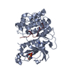
Spodoptera frugiperda (fall armyworm) / References: UniProt: Q62074, protein kinase C
#2: Protein/peptide Partitioning defective 3 homolog / PAR-3 / PARD-3 / Atypical PKC isotype-specific-interacting protein / ASIP / Atypical PKC-specific- ...PAR-3 / PARD-3 / Atypical PKC isotype-specific-interacting protein / ASIP / Atypical PKC-specific-binding protein / ASBP


Mass: 3207.556 Da / Num. of mol.: 1 / Fragment: Substrate Peptide, UNP residues 813-840 / Source method: obtained synthetically
Details: This sequence occurs naturally in humans/mouses/rats.
Source: (synth.) Rattus norvegicus (Norway rat) / References: UniProt: Q9Z340
#3: Chemical ChemComp-ADE / ADENINE / Adenine


Mass: 135.127 Da / Num. of mol.: 1 / Source method: obtained synthetically / Formula: C5H5N5
#4: Water ChemComp-HOH / water / Water


Mass: 18.015 Da / Num. of mol.: 52 / Source method: isolated from a natural source / Formula: H2O

-
Experimental details

-
Experiment

ExperimentMethod: X-RAY DIFFRACTION / Number of used crystals: 1

-
Sample preparation

CrystalDensity Matthews: 2.13 Å3/Da / Density % sol: 42.13 %
Crystal growTemperature: 289 K / Method: vapor diffusion, hanging drop / pH: 7
Details: 3.5M sodium formate, pH 7.0, VAPOR DIFFUSION, HANGING DROP, temperature 289K

-
Data collection

DiffractionMean temperature: 200 K
Diffraction sourceSource: SYNCHROTRON / Site: SSRF / Beamline: BL17U
DetectorType: RAYONIX MX-225 / Detector: CCD / Date: Nov 6, 2010
RadiationProtocol: SINGLE WAVELENGTH / Monochromatic (M) / Laue (L): M / Scattering type: x-ray
Radiation wavelengthRelative weight: 1
ReflectionResolution: 2.4→21.2 Å / Num. all: 16230 / Num. obs: 13400 / % possible obs: 82.6 % / Observed criterion σ(F): 0 / Observed criterion σ(I): 2 / Biso Wilson estimate: 32.72 Å2

-
Processing

Software
NameVersionClassification
HKL-2000data collection
PHASESphasing
PHENIX(phenix.refine: 1.7_629)refinement
HKL-2000data reduction
HKL-2000data scaling
RefinementMethod to determine structure: MOLECULAR REPLACEMENT / Resolution: 2.4→21.179 Å / Occupancy max: 1 / Occupancy min: 0 / FOM work R set: 0.8302 / SU ML: 0.33 / σ(F): 0 / Phase error: 24.25 / Stereochemistry target values: ML
RfactorNum. reflection% reflectionSelection details
Rfree0.2414 659 4.92 %RANDOM
Rwork0.1723 ---
all0.1757 16230 --
obs0.1757 13400 82.56 %-
Solvent computationShrinkage radii: 0.83 Å / VDW probe radii: 1.1 Å / Solvent model: FLAT BULK SOLVENT MODEL / Bsol: 45.251 Å2 / ksol: 0.344 e/Å3
Displacement parametersBiso max: 178.12 Å2 / Biso mean: 43.3645 Å2 / Biso min: 6.88 Å2
Baniso -1Baniso -2Baniso -3
1--2.1692 Å2-0 Å21.2238 Å2
2--0.697 Å2-0 Å2
3---1.4722 Å2
Refinement stepCycle: LAST / Resolution: 2.4→21.179 Å
ProteinNucleic acidLigandSolventTotal
Num. atoms2683 0 10 52 2745
Refine LS restraints
Refine-IDTypeDev idealNumber
X-RAY DIFFRACTIONf_bond_d0.0082764
X-RAY DIFFRACTIONf_angle_d1.1323747
X-RAY DIFFRACTIONf_chiral_restr0.078406
X-RAY DIFFRACTIONf_plane_restr0.005485
X-RAY DIFFRACTIONf_dihedral_angle_d14.204981
LS refinement shell

Refine-ID: X-RAY DIFFRACTION / Total num. of bins used: 5

Resolution (Å)Rfactor RfreeNum. reflection RfreeRfactor RworkNum. reflection RworkNum. reflection all% reflection obs (%)
2.4-2.5850.2991190.21362321244076
2.585-2.84460.29821290.20482509263882
2.8446-3.2550.27231320.19042647277986
3.255-4.09610.24881300.15562651278186
4.0961-21.18030.19731490.15562613276283
Refinement TLS params.

Method: refined / Refine-ID: X-RAY DIFFRACTION

IDL112)L122)L132)L222)L232)L332)S11 (Å °)S12 (Å °)S13 (Å °)S21 (Å °)S22 (Å °)S23 (Å °)S31 (Å °)S32 (Å °)S33 (Å °)T112)T122)T132)T222)T232)T332)Origin x (Å)Origin y (Å)Origin z (Å)
10.52960.38860.43060.5216-0.04530.762-0.13340.2568-0.0035-0.12770.1525-0.0873-0.19560.0688-0.03550.1875-0.04660.03480.18020.00110.12936.5796-9.711313.7984
20.24940.01050.04670.0624-0.13170.5965-0.009-0.0222-0.16270.06650.0475-0.15270.2727-0.1993-0.04730.09930.0122-0.05550.03950.00980.15092.4416-25.608827.4723
30.25850.03370.09790.36410.05810.67010.0551-0.0014-0.01010.0130.41920.1662-0.4131-0.54950.06410.0402-0.1655-0.0554-0.1354-0.2453-0.07950.0215-12.838119.0586
40.2002-0.02060.10390.62550.0760.08550.09540.0209-0.09740.09090.0117-0.25220.1370.0877-0.00240.1578-0.05110.02530.25440.01360.35179.9009-15.103731.7255
Refinement TLS group
IDRefine-IDRefine TLS-IDSelection detailsAuth asym-IDAuth seq-ID
1X-RAY DIFFRACTION1(chain A and resid 239:394)A239 - 394
2X-RAY DIFFRACTION2(chain A and resid 395:469)A395 - 469
3X-RAY DIFFRACTION3(chain A and resid 470:577)A470 - 577
4X-RAY DIFFRACTION4(chain Z and resid 1047:1062)Z1047 - 1062

+
About Yorodumi

-
News

-
Feb 9, 2022. New format data for meta-information of EMDB entries

New format data for meta-information of EMDB entries

  • Version 3 of the EMDB header file is now the official format.
  • The previous official version 1.9 will be removed from the archive.

Related info.:EMDB header

External links:wwPDB to switch to version 3 of the EMDB data model

-
Aug 12, 2020. Covid-19 info

Covid-19 info

URL: https://pdbj.org/emnavi/covid19.php

New page: Covid-19 featured information page in EM Navigator.

Related info.:Covid-19 info / Mar 5, 2020. Novel coronavirus structure data

+
Mar 5, 2020. Novel coronavirus structure data

Novel coronavirus structure data

Related info.:Yorodumi Speices / Aug 12, 2020. Covid-19 info

External links:COVID-19 featured content - PDBj / Molecule of the Month (242):Coronavirus Proteases

+
Jan 31, 2019. EMDB accession codes are about to change! (news from PDBe EMDB page)

EMDB accession codes are about to change! (news from PDBe EMDB page)

  • The allocation of 4 digits for EMDB accession codes will soon come to an end. Whilst these codes will remain in use, new EMDB accession codes will include an additional digit and will expand incrementally as the available range of codes is exhausted. The current 4-digit format prefixed with “EMD-” (i.e. EMD-XXXX) will advance to a 5-digit format (i.e. EMD-XXXXX), and so on. It is currently estimated that the 4-digit codes will be depleted around Spring 2019, at which point the 5-digit format will come into force.
  • The EM Navigator/Yorodumi systems omit the EMD- prefix.

Related info.:Q: What is EMD? / ID/Accession-code notation in Yorodumi/EM Navigator

External links:EMDB Accession Codes are Changing Soon! / Contact to PDBj

+
Jul 12, 2017. Major update of PDB

Major update of PDB

  • wwPDB released updated PDB data conforming to the new PDBx/mmCIF dictionary.
  • This is a major update changing the version number from 4 to 5, and with Remediation, in which all the entries are updated.
  • In this update, many items about electron microscopy experimental information are reorganized (e.g. em_software).
  • Now, EM Navigator and Yorodumi are based on the updated data.

External links:wwPDB Remediation / Enriched Model Files Conforming to OneDep Data Standards Now Available in the PDB FTP Archive

-
Yorodumi

Thousand views of thousand structures

  • Yorodumi is a browser for structure data from EMDB, PDB, SASBDB, etc.
  • This page is also the successor to EM Navigator detail page, and also detail information page/front-end page for Omokage search.
  • The word "yorodu" (or yorozu) is an old Japanese word meaning "ten thousand". "mi" (miru) is to see.

Related info.:EMDB / PDB / SASBDB / Comparison of 3 databanks / Yorodumi Search / Aug 31, 2016. New EM Navigator & Yorodumi / Yorodumi Papers / Jmol/JSmol / Function and homology information / Changes in new EM Navigator and Yorodumi

Read more