 Movie
Movie Controller

[English] 日本語
 Yorodumi
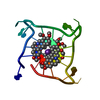
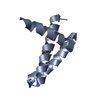
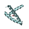
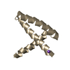
- PDB-4da3: Crystal structure of an intramolecular human telomeric DNA G-quad... -
+ Open data

- Basic information
| Entry | Database: PDB / ID: 4da3 | ||||||||||||||||||||
|---|---|---|---|---|---|---|---|---|---|---|---|---|---|---|---|---|---|---|---|---|---|
| Title | Crystal structure of an intramolecular human telomeric DNA G-quadruplex 21-mer bound by the naphthalene diimide compound MM41. | ||||||||||||||||||||
 Components Components | DNA (5'-D(* Keywords KeywordsDNA / intramolecular / ligand-complex / G-quadruplex / telomere | Function / homology | Chem-0DX / : / DNA / DNA (> 10) |  Function and homology information Function and homology informationBiological species | Synthetic DNA (others) | Method |  X-RAY DIFFRACTION / SYNCHROTRON / MOLECULAR REPLACEMENT / Resolution: 2.4 Å X-RAY DIFFRACTION / SYNCHROTRON / MOLECULAR REPLACEMENT / Resolution: 2.4 Å  Authors AuthorsCollie, G.W. / Neidle, S. |  CitationJournal: J.Med.Chem. / Year: 2013 CitationJournal: J.Med.Chem. / Year: 2013Title: Structure-based design and evaluation of naphthalene diimide g-quadruplex ligands as telomere targeting agents in pancreatic cancer cells. Authors: Micco, M. / Collie, G.W. / Dale, A.G. / Ohnmacht, S.A. / Pazitna, I. / Gunaratnam, M. / Reszka, A.P. / Neidle, S. History |
|
- Structure visualization
| Structure viewer | Molecule: MolmilJmol/JSmol |
|---|
- Downloads & links
-Download
| PDBx/mmCIF format | 4da3.cif.gz | 24.6 KB | Display | PDBx/mmCIF format |
|---|---|---|---|---|
| PDB format | pdb4da3.ent.gz | 16.9 KB | Display | PDB format |
| PDBx/mmJSON format | 4da3.json.gz | Tree view | PDBx/mmJSON format | |
| Others |  Other downloads Other downloads |
-Validation report
| Arichive directory | https://data.pdbj.org/pub/pdb/validation_reports/da/4da3ftp://data.pdbj.org/pub/pdb/validation_reports/da/4da3 | HTTPS FTP |
|---|
-Related structure data
| Related structure data |  3uyhC  4daqC  3sc8S C: citing same article ( S: Starting model for refinement |
|---|---|
| Similar structure data |

-Links
 PDBj
PDBj


- Assembly
| Deposited unit | 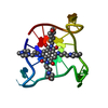
| ||||||||
|---|---|---|---|---|---|---|---|---|---|
| 1 |
| ||||||||
| Unit cell |
| ||||||||
| Components on special symmetry positions |
|
-Components
| #1: DNA chain | Mass: 6670.290 Da / Num. of mol.: 1 / Source method: obtained synthetically Details: DNA synthesized by standard phosphoramidite chemistry. Source: (synth.) Synthetic DNA (others) | ||||
|---|---|---|---|---|---|
| #2: Chemical |   Mass: 39.098 Da / Num. of mol.: 3 / Source method: obtained synthetically / Formula: K Mass: 39.098 Da / Num. of mol.: 3 / Source method: obtained synthetically / Formula: K#3: Chemical | ChemComp-0DX / | 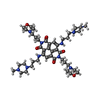  Mass: 831.058 Da / Num. of mol.: 1 / Source method: obtained synthetically / Formula: C44H66N10O6 Mass: 831.058 Da / Num. of mol.: 1 / Source method: obtained synthetically / Formula: C44H66N10O6#4: Water | ChemComp-HOH / |  Mass: 18.015 Da / Num. of mol.: 25 / Source method: isolated from a natural source / Formula: H2O Mass: 18.015 Da / Num. of mol.: 25 / Source method: isolated from a natural source / Formula: H2O |
-Experimental details
-Experiment
| Experiment | Method: X-RAY DIFFRACTION / Number of used crystals: 1 |
|---|
- Sample preparation
| Crystal | Density Matthews: 2.96 Å3/Da / Density % sol: 58.43 % |
|---|---|
| Crystal grow | Temperature: 293 K / Method: vapor diffusion, hanging drop / pH: 6.5 Details: 15% PEG400, 150 mM sodium chloride and 50 mM sodium cacodylate (pH 6.5), VAPOR DIFFUSION, HANGING DROP, temperature 293K |
-Data collection
| Diffraction | Mean temperature: 100 K |
|---|---|
| Diffraction source | Source: SYNCHROTRON / Site: Diamond  / Beamline: I04 / Wavelength: 0.9795 Å / Beamline: I04 / Wavelength: 0.9795 Å |
| Detector | Type: ADSC QUANTUM 315r / Detector: CCD / Date: Dec 16, 2011 |
| Radiation | Monochromator: SI 111 CHANNEL / Protocol: SINGLE WAVELENGTH / Monochromatic (M) / Laue (L): M / Scattering type: x-ray |
| Radiation wavelength | Wavelength: 0.9795 Å / Relative weight: 1 |
| Reflection | Resolution: 2.27→25.97 Å / Num. all: 3843 / Num. obs: 3843 / % possible obs: 98.9 % / Observed criterion σ(F): 2 / Observed criterion σ(I): 2 / Redundancy: 6.7 % / Biso Wilson estimate: 66.213 Å2 / Rmerge(I) obs: 0.038 / Net I/σ(I): 23.9 |
| Reflection shell | Resolution: 2.27→2.33 Å / Redundancy: 5.8 % / Rmerge(I) obs: 0.597 / Mean I/σ(I) obs: 2.3 / Num. unique all: 278 / % possible all: 100 |
- Processing
| Software |
| |||||||||||||||||||||||||
|---|---|---|---|---|---|---|---|---|---|---|---|---|---|---|---|---|---|---|---|---|---|---|---|---|---|---|
| Refinement | Method to determine structure: MOLECULAR REPLACEMENT Starting model: PDB ENTRY 3SC8 Resolution: 2.4→14.81 Å / Cor.coef. Fo:Fc: 0.939 / Cor.coef. Fo:Fc free: 0.921 / SU B: 10.523 / SU ML: 0.226 / Cross valid method: THROUGHOUT / ESU R: 0.441 / ESU R Free: 0.287 / Stereochemistry target values: MAXIMUM LIKELIHOOD / Details: HYDROGENS HAVE BEEN USED IF PRESENT IN THE INPUT
| |||||||||||||||||||||||||
| Solvent computation | Ion probe radii: 0.8 Å / Shrinkage radii: 0.8 Å / VDW probe radii: 1.2 Å / Solvent model: MASK | |||||||||||||||||||||||||
| Displacement parameters | Biso mean: 39.532 Å2
| |||||||||||||||||||||||||
| Refinement step | Cycle: LAST / Resolution: 2.4→14.81 Å
| |||||||||||||||||||||||||
| Refine LS restraints |
| |||||||||||||||||||||||||
| LS refinement shell | Resolution: 2.4→2.461 Å / Total num. of bins used: 20
|
