[English] 日本語
Yorodumi
- PDB-4d07: DYNLL2 dynein light chain binds to an extended, unstructured line... -

+
Open data


ID or keywords:

Loading...

-
Basic information

Entry
Database: PDB / ID: 4d07
Title
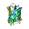
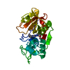
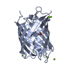
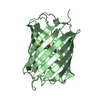
DYNLL2 dynein light chain binds to an extended, unstructured linear motif of myosin 5a tail
Components
  • DYNEIN LIGHT CHAIN 2, CYTOPLASMIC
  • MYOSIN VA VARIANT
KeywordsMOTOR PROTEIN / INSTRINSICALLY DISORDERED DOMAIN / HUB PROTEIN
Function / homology
Function and homology information


myosin V complex / Activation of BMF and translocation to mitochondria / 9+0 non-motile cilium / post-Golgi vesicle-mediated transport / insulin-responsive compartment / melanosome transport / actin filament-based movement / ciliary tip / filopodium tip / Intraflagellar transport ...myosin V complex / Activation of BMF and translocation to mitochondria / 9+0 non-motile cilium / post-Golgi vesicle-mediated transport / insulin-responsive compartment / melanosome transport / actin filament-based movement / ciliary tip / filopodium tip / Intraflagellar transport / COPI-independent Golgi-to-ER retrograde traffic / cytoplasmic dynein complex / vesicle transport along actin filament / myosin complex / microfilament motor activity / Macroautophagy / dynein intermediate chain binding / Insulin processing / cytoskeletal motor activity / Amplification of signal from unattached kinetochores via a MAD2 inhibitory signal / microtubule-based process / COPI-mediated anterograde transport / Mitotic Prometaphase / EML4 and NUDC in mitotic spindle formation / vesicle-mediated transport / ruffle / Resolution of Sister Chromatid Cohesion / HSP90 chaperone cycle for steroid hormone receptors (SHR) in the presence of ligand / MHC class II antigen presentation / actin filament organization / RHO GTPases Activate Formins / Translocation of SLC2A4 (GLUT4) to the plasma membrane / protein localization to plasma membrane / FCGR3A-mediated phagocytosis / cilium / Regulation of actin dynamics for phagocytic cup formation / small GTPase binding / Aggrephagy / HCMV Early Events / cellular response to insulin stimulus / Separation of Sister Chromatids / melanosome / actin filament binding / actin cytoskeleton / protein transport / actin binding / growth cone / postsynapse / microtubule / cytoskeleton / calmodulin binding / neuron projection / centrosome / glutamatergic synapse / RNA binding / extracellular exosome / ATP binding / membrane / nucleus / plasma membrane / cytosol / cytoplasm
Similarity search - Function
Protein Inhibitor Of Neuronal Nitric Oxide Synthase / Protein Inhibitor Of Neuronal Nitric Oxide Synthase; / Dynein light chain, type 1/2, conserved site / Dynein light chain type 1 signature. / Myosin 5a, cargo-binding domain / Dynein light chain type 1 / Dynein light chain, type 1/2 / Dynein light chain superfamily / Dynein light chain type 1 / Class V myosin, motor domain ...Protein Inhibitor Of Neuronal Nitric Oxide Synthase / Protein Inhibitor Of Neuronal Nitric Oxide Synthase; / Dynein light chain, type 1/2, conserved site / Dynein light chain type 1 signature. / Myosin 5a, cargo-binding domain / Dynein light chain type 1 / Dynein light chain, type 1/2 / Dynein light chain superfamily / Dynein light chain type 1 / Class V myosin, motor domain / Dilute domain / DIL domain / Dilute domain profile. / DIL / IQ calmodulin-binding motif / Myosin, N-terminal, SH3-like / Myosin N-terminal SH3-like domain profile. / Short calmodulin-binding motif containing conserved Ile and Gln residues. / Myosin head, motor domain / Myosin head (motor domain) / Myosin motor domain profile. / Myosin. Large ATPases. / IQ motif profile. / IQ motif, EF-hand binding site / Kinesin motor domain superfamily / P-loop containing nucleoside triphosphate hydrolase / 2-Layer Sandwich / Alpha Beta
Similarity search - Domain/homology
: / Myosin Va variant / Dynein light chain 2, cytoplasmic / Unconventional myosin-Va
Similarity search - Component
Biological speciesHOMO SAPIENS (human)
MethodX-RAY DIFFRACTION / SYNCHROTRON / MOLECULAR REPLACEMENT / Resolution: 1.85 Å
AuthorsBodor, A. / Radnai, L. / Hetenyi, C. / Rapali, P. / Lang, A. / Kover, K.E. / Perczel, A. / Wahlgren, W.Y. / Katona, G. / Nyitray, L.
CitationJournal: Biochemistry / Year: 2014
Title: Dynll2 Dynein Light Chain Binds to an Extended Linear Motif of Myosin 5A Tail that Has Structural Plasticity.
Authors: Bodor, A. / Radnai, L. / Hetenyi, C. / Rapali, P. / Lang, A. / Kover, K.E. / Perczel, A. / Wahlgren, W.Y. / Katona, G. / Nyitray, L.
History
DepositionApr 24, 2014Deposition site: PDBE / Processing site: PDBE
Revision 1.0Oct 22, 2014Provider: repository / Type: Initial release
Revision 1.1Oct 29, 2014Group: Database references
Revision 1.2Dec 3, 2014Group: Database references
Revision 1.3Dec 20, 2023Group: Data collection / Database references ...Data collection / Database references / Derived calculations / Other / Refinement description
Category: chem_comp_atom / chem_comp_bond ...chem_comp_atom / chem_comp_bond / database_2 / pdbx_database_status / pdbx_initial_refinement_model / pdbx_struct_conn_angle / struct_conn / struct_site
Item: _database_2.pdbx_DOI / _database_2.pdbx_database_accession ..._database_2.pdbx_DOI / _database_2.pdbx_database_accession / _pdbx_database_status.status_code_sf / _pdbx_struct_conn_angle.ptnr1_auth_comp_id / _pdbx_struct_conn_angle.ptnr1_auth_seq_id / _pdbx_struct_conn_angle.ptnr1_label_atom_id / _pdbx_struct_conn_angle.ptnr1_label_comp_id / _pdbx_struct_conn_angle.ptnr1_label_seq_id / _pdbx_struct_conn_angle.ptnr3_auth_comp_id / _pdbx_struct_conn_angle.ptnr3_auth_seq_id / _pdbx_struct_conn_angle.ptnr3_label_atom_id / _pdbx_struct_conn_angle.ptnr3_label_comp_id / _pdbx_struct_conn_angle.ptnr3_label_seq_id / _pdbx_struct_conn_angle.value / _struct_conn.pdbx_dist_value / _struct_conn.ptnr1_auth_comp_id / _struct_conn.ptnr1_auth_seq_id / _struct_conn.ptnr1_label_asym_id / _struct_conn.ptnr1_label_atom_id / _struct_conn.ptnr1_label_comp_id / _struct_conn.ptnr1_label_seq_id / _struct_conn.ptnr2_auth_comp_id / _struct_conn.ptnr2_auth_seq_id / _struct_conn.ptnr2_label_asym_id / _struct_conn.ptnr2_label_atom_id / _struct_conn.ptnr2_label_comp_id / _struct_conn.ptnr2_label_seq_id / _struct_site.pdbx_auth_asym_id / _struct_site.pdbx_auth_comp_id / _struct_site.pdbx_auth_seq_id

-
Structure visualization

Structure viewerMolecule:
MolmilJmol/JSmol

Downloads & links

-
Assembly

Deposited unit
A: DYNEIN LIGHT CHAIN 2, CYTOPLASMIC
B: MYOSIN VA VARIANT
hetero molecules


Theoretical massNumber of molelcules
Total (without water)13,9475
Polymers13,6692
Non-polymers2773
Water1,78399
1
A: DYNEIN LIGHT CHAIN 2, CYTOPLASMIC
B: MYOSIN VA VARIANT
hetero molecules

A: DYNEIN LIGHT CHAIN 2, CYTOPLASMIC
B: MYOSIN VA VARIANT
hetero molecules


Theoretical massNumber of molelcules
Total (without water)27,89310
Polymers27,3394
Non-polymers5546
Water724
TypeNameSymmetry operationNumber
identity operation1_555x,y,z1
crystal symmetry operation10_774-y+2,-x+2,-z-1/61
Buried area6140 Å2
ΔGint-62.8 kcal/mol
Surface area9160 Å2
MethodPISA
Unit cell
Length a, b, c (Å)45.160, 45.160, 204.010
Angle α, β, γ (deg.)90.00, 90.00, 120.00
Int Tables number178
Space group name H-MP6122
Components on special symmetry positions
IDModelComponents
11A-2051-

HOH

-
Components

-
Protein / Protein/peptide , 2 types, 2 molecules AB

#1: Protein DYNEIN LIGHT CHAIN 2, CYTOPLASMIC / 8 KDA DYNEIN LIGHT CHAIN B / DLC8B / DYNEIN LIGHT CHAIN LC8-TYPE 2 / DYNLL2


Mass: 10647.124 Da / Num. of mol.: 1
Source method: isolated from a genetically manipulated source
Source: (gene. exp.) HOMO SAPIENS (human) / Production host: ESCHERICHIA COLI (E. coli) / Strain (production host): BL21(DE3) / Variant (production host): ROSETTA / References: UniProt: Q96FJ2
#2: Protein/peptide MYOSIN VA VARIANT / MYO5A


Mass: 3022.365 Da / Num. of mol.: 1 / Fragment: RESIDUES 856-878 / Source method: obtained synthetically / Source: (synth.) HOMO SAPIENS (human) / References: UniProt: Q59FF5, UniProt: Q9Y4I1*PLUS

-
Non-polymers , 4 types, 102 molecules

#3: Chemical ChemComp-CO / COBALT (II) ION


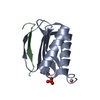
Mass: 58.933 Da / Num. of mol.: 1 / Source method: obtained synthetically / Formula: Co
#4: Chemical ChemComp-TRS / 2-AMINO-2-HYDROXYMETHYL-PROPANE-1,3-DIOL / TRIS BUFFER / Tris


Mass: 122.143 Da / Num. of mol.: 1 / Source method: obtained synthetically / Formula: C4H12NO3 / Comment: pH buffer*YM
#5: Chemical ChemComp-SO4 / SULFATE ION / Sulfate


Mass: 96.063 Da / Num. of mol.: 1 / Source method: obtained synthetically / Formula: SO4
#6: Water ChemComp-HOH / water / Water


Mass: 18.015 Da / Num. of mol.: 99 / Source method: isolated from a natural source / Formula: H2O

-
Experimental details

-
Experiment

ExperimentMethod: X-RAY DIFFRACTION / Number of used crystals: 1

-
Sample preparation

CrystalDensity Matthews: 2.51 Å3/Da / Density % sol: 51.03 % / Description: NONE
Crystal growDetails: 2.5UL PROTEIN PLUS 1UL RESERVOIR SOLUTION OF 1.8 M (NH4)2SO4, 0.02 M COCL2, 0.1 M MES PH 6.5

-
Data collection

DiffractionMean temperature: 100 K
Diffraction sourceSource: SYNCHROTRON / Site: ESRF / Beamline: ID29 / Wavelength: 0.93
DetectorType: ADSC QUANTUM 315r / Detector: CCD / Date: Nov 9, 2003
RadiationProtocol: SINGLE WAVELENGTH / Monochromatic (M) / Laue (L): M / Scattering type: x-ray
Radiation wavelengthWavelength: 0.93 Å / Relative weight: 1
ReflectionResolution: 1.85→19.76 Å / Num. obs: 11478 / % possible obs: 99.8 % / Observed criterion σ(I): 2 / Redundancy: 22 % / Rmerge(I) obs: 0.1 / Net I/σ(I): 30.18
Reflection shellResolution: 1.85→1.9 Å / Redundancy: 20 % / Rmerge(I) obs: 0.7 / Mean I/σ(I) obs: 6.09 / % possible all: 100

-
Processing

Software
NameVersionClassification
REFMAC5.8.0069refinement
XDSdata reduction
XSCALEdata scaling
PHASERphasing
RefinementMethod to determine structure: MOLECULAR REPLACEMENT
Starting model: PDB ENTRY 1CMI
Resolution: 1.85→204.01 Å / Cor.coef. Fo:Fc: 0.954 / Cor.coef. Fo:Fc free: 0.93 / SU B: 2.97 / SU ML: 0.091 / Cross valid method: THROUGHOUT / ESU R: 0.138 / ESU R Free: 0.133 / Stereochemistry target values: MAXIMUM LIKELIHOOD / Details: HYDROGENS HAVE BEEN ADDED IN THE RIDING POSITIONS.
RfactorNum. reflection% reflectionSelection details
Rfree0.233 544 4.8 %RANDOM
Rwork0.19303 ---
obs0.19492 10853 99.82 %-
Solvent computationIon probe radii: 0.8 Å / Shrinkage radii: 0.8 Å / VDW probe radii: 1.2 Å / Solvent model: MASK
Displacement parametersBiso mean: 21.851 Å2
Baniso -1Baniso -2Baniso -3
1-0.75 Å20.37 Å20 Å2
2--0.75 Å20 Å2
3----2.42 Å2
Refinement stepCycle: LAST / Resolution: 1.85→204.01 Å
ProteinNucleic acidLigandSolventTotal
Num. atoms841 0 14 99 954
Refine LS restraints
Refine-IDTypeDev idealDev ideal targetNumber
X-RAY DIFFRACTIONr_bond_refined_d0.0070.019918
X-RAY DIFFRACTIONr_bond_other_d
X-RAY DIFFRACTIONr_angle_refined_deg1.1131.9411250
X-RAY DIFFRACTIONr_angle_other_deg
X-RAY DIFFRACTIONr_dihedral_angle_1_deg5.5485121
X-RAY DIFFRACTIONr_dihedral_angle_2_deg31.12625.68244
X-RAY DIFFRACTIONr_dihedral_angle_3_deg13.28915169
X-RAY DIFFRACTIONr_dihedral_angle_4_deg0.183151
X-RAY DIFFRACTIONr_chiral_restr0.0860.2134
X-RAY DIFFRACTIONr_gen_planes_refined0.0040.02696
X-RAY DIFFRACTIONr_gen_planes_other
X-RAY DIFFRACTIONr_nbd_refined
X-RAY DIFFRACTIONr_nbd_other
X-RAY DIFFRACTIONr_nbtor_refined
X-RAY DIFFRACTIONr_nbtor_other
X-RAY DIFFRACTIONr_xyhbond_nbd_refined
X-RAY DIFFRACTIONr_xyhbond_nbd_other
X-RAY DIFFRACTIONr_metal_ion_refined
X-RAY DIFFRACTIONr_metal_ion_other
X-RAY DIFFRACTIONr_symmetry_vdw_refined
X-RAY DIFFRACTIONr_symmetry_vdw_other
X-RAY DIFFRACTIONr_symmetry_hbond_refined
X-RAY DIFFRACTIONr_symmetry_hbond_other
X-RAY DIFFRACTIONr_symmetry_metal_ion_refined
X-RAY DIFFRACTIONr_symmetry_metal_ion_other
X-RAY DIFFRACTIONr_mcbond_it1.0461.898438
X-RAY DIFFRACTIONr_mcbond_other
X-RAY DIFFRACTIONr_mcangle_it1.8052.807549
X-RAY DIFFRACTIONr_mcangle_other
X-RAY DIFFRACTIONr_scbond_it1.2772.116479
X-RAY DIFFRACTIONr_scbond_other
X-RAY DIFFRACTIONr_scangle_it
X-RAY DIFFRACTIONr_scangle_other
X-RAY DIFFRACTIONr_long_range_B_refined
X-RAY DIFFRACTIONr_long_range_B_other
X-RAY DIFFRACTIONr_rigid_bond_restr
X-RAY DIFFRACTIONr_sphericity_free
X-RAY DIFFRACTIONr_sphericity_bonded
LS refinement shellResolution: 1.85→1.898 Å / Total num. of bins used: 20
RfactorNum. reflection% reflection
Rfree0.313 34 -
Rwork0.251 788 -
obs--99.64 %

+
About Yorodumi

-
News

-
Feb 9, 2022. New format data for meta-information of EMDB entries

New format data for meta-information of EMDB entries

  • Version 3 of the EMDB header file is now the official format.
  • The previous official version 1.9 will be removed from the archive.

Related info.:EMDB header

External links:wwPDB to switch to version 3 of the EMDB data model

-
Aug 12, 2020. Covid-19 info

Covid-19 info

URL: https://pdbj.org/emnavi/covid19.php

New page: Covid-19 featured information page in EM Navigator.

Related info.:Covid-19 info / Mar 5, 2020. Novel coronavirus structure data

+
Mar 5, 2020. Novel coronavirus structure data

Novel coronavirus structure data

Related info.:Yorodumi Speices / Aug 12, 2020. Covid-19 info

External links:COVID-19 featured content - PDBj / Molecule of the Month (242):Coronavirus Proteases

+
Jan 31, 2019. EMDB accession codes are about to change! (news from PDBe EMDB page)

EMDB accession codes are about to change! (news from PDBe EMDB page)

  • The allocation of 4 digits for EMDB accession codes will soon come to an end. Whilst these codes will remain in use, new EMDB accession codes will include an additional digit and will expand incrementally as the available range of codes is exhausted. The current 4-digit format prefixed with “EMD-” (i.e. EMD-XXXX) will advance to a 5-digit format (i.e. EMD-XXXXX), and so on. It is currently estimated that the 4-digit codes will be depleted around Spring 2019, at which point the 5-digit format will come into force.
  • The EM Navigator/Yorodumi systems omit the EMD- prefix.

Related info.:Q: What is EMD? / ID/Accession-code notation in Yorodumi/EM Navigator

External links:EMDB Accession Codes are Changing Soon! / Contact to PDBj

+
Jul 12, 2017. Major update of PDB

Major update of PDB

  • wwPDB released updated PDB data conforming to the new PDBx/mmCIF dictionary.
  • This is a major update changing the version number from 4 to 5, and with Remediation, in which all the entries are updated.
  • In this update, many items about electron microscopy experimental information are reorganized (e.g. em_software).
  • Now, EM Navigator and Yorodumi are based on the updated data.

External links:wwPDB Remediation / Enriched Model Files Conforming to OneDep Data Standards Now Available in the PDB FTP Archive

-
Yorodumi

Thousand views of thousand structures

  • Yorodumi is a browser for structure data from EMDB, PDB, SASBDB, etc.
  • This page is also the successor to EM Navigator detail page, and also detail information page/front-end page for Omokage search.
  • The word "yorodu" (or yorozu) is an old Japanese word meaning "ten thousand". "mi" (miru) is to see.

Related info.:EMDB / PDB / SASBDB / Comparison of 3 databanks / Yorodumi Search / Aug 31, 2016. New EM Navigator & Yorodumi / Yorodumi Papers / Jmol/JSmol / Function and homology information / Changes in new EM Navigator and Yorodumi

Read more