 Movie
Movie Controller

[English] 日本語
 Yorodumi
- PDB-4cwe: Structural studies of rolling circle replication initiation prote... -
+ Open data

- Basic information
| Entry | Database: PDB / ID: 4cwe | ||||||
|---|---|---|---|---|---|---|---|
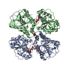
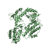
| Title | Structural studies of rolling circle replication initiation protein from Staphylococcus aureus | ||||||
 Components Components | REPLICATION INITIATION PROTEIN, REPLICATION INITIATION PROTEIN | ||||||
 Keywords Keywords | ISOMERASE / ANTIBIOTIC RESISTANCE / PCRA HELICASE / DNA RELAXASE / CHIMERA | ||||||
| Function / homology | Replication initiation factor / : / Replication initiation factor / Replication initiation protein, N-terminal / DNA replication / Replication initiation protein / Replication initiation protein Function and homology information Function and homology information | ||||||
| Biological species |   STAPHYLOCOCCUS AUREUS (bacteria) STAPHYLOCOCCUS AUREUS (bacteria) | ||||||
| Method |  X-RAY DIFFRACTION / SYNCHROTRON / MOLECULAR REPLACEMENT / Resolution: 3 Å X-RAY DIFFRACTION / SYNCHROTRON / MOLECULAR REPLACEMENT / Resolution: 3 Å | ||||||
 Authors Authors | Carr, S.B. / Phillips, S.E.V. / Thomas, C.D. | ||||||
 Citation Citation | Journal: Nucleic Acids Res. / Year: 2016 Title: Structures of Replication Initiation Proteins from Staphylococcal Antibiotic Resistance Plasmids Reveal Protein Asymmetry and Flexibility are Necessary for Replication. Authors: Carr, S.B. / Phillips, S.E. / Thomas, C.D. | ||||||
| History |
|
- Structure visualization
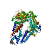
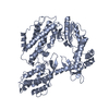
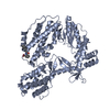
| Structure viewer | Molecule: MolmilJmol/JSmol |
|---|
- Downloads & links
-Download
| PDBx/mmCIF format | 4cwe.cif.gz | 124.8 KB | Display | PDBx/mmCIF format |
|---|---|---|---|---|
| PDB format | pdb4cwe.ent.gz | 99.1 KB | Display | PDB format |
| PDBx/mmJSON format | 4cwe.json.gz | Tree view | PDBx/mmJSON format | |
| Others |  Other downloads Other downloads |
-Validation report
| Arichive directory | https://data.pdbj.org/pub/pdb/validation_reports/cw/4cweftp://data.pdbj.org/pub/pdb/validation_reports/cw/4cwe | HTTPS FTP |
|---|
-Related structure data
| Related structure data | 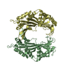 4cijSC  4cwcC C: citing same article ( S: Starting model for refinement |
|---|---|
| Similar structure data |

-Links
 PDBj
PDBj- Assembly
| Deposited unit | 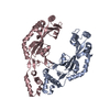
| ||||||||
|---|---|---|---|---|---|---|---|---|---|
| 1 |
| ||||||||
| Unit cell |
|
-Components
| #1: Protein | Mass: 34236.105 Da / Num. of mol.: 2 Fragment: REPLICATION INITIATION PROTEIN RESIDUES 32-216, REPLICATION INITIATION PROTEIN RESIDUES 220-314 Source method: isolated from a genetically manipulated source Source: (gene. exp.) STAPHYLOCOCCUS AUREUS (bacteria) / Plasmid: PET-15M / Production host: Sequence details | PROTEIN WAS CONSTRUCTED BY FUSING THE N-TERMINAL DOMAIN OF REPD - BASES 1295-1849 FROM PLASMID ...PROTEIN WAS CONSTRUCTE | |
|---|
-Experimental details
-Experiment
| Experiment | Method: X-RAY DIFFRACTION / Number of used crystals: 1 |
|---|
- Sample preparation
| Crystal | Density Matthews: 2.92 Å3/Da / Density % sol: 57.8 % / Description: NONE |
|---|---|
| Crystal grow | pH: 5.5 / Details: 0.1 M SODIUM CITRATE PH 5.5, 2.5 M 1,6-HEXANEDIOL |
-Data collection
| Diffraction | Mean temperature: 100 K | |||||||||||||||
|---|---|---|---|---|---|---|---|---|---|---|---|---|---|---|---|---|
| Diffraction source | Source: SYNCHROTRON / Site: SRS  / Beamline: PX14.1 / Wavelength: 1.488 / Beamline: PX14.1 / Wavelength: 1.488 | |||||||||||||||
| Detector | Type: ADSC Q4 / Detector: CCD / Date: Jun 7, 2002 / Details: MIRRORS | |||||||||||||||
| Radiation | Protocol: SINGLE WAVELENGTH / Monochromatic (M) / Laue (L): M / Scattering type: x-ray | |||||||||||||||
| Radiation wavelength | Wavelength: 1.488 Å / Relative weight: 1 | |||||||||||||||
| Reflection twin |
| |||||||||||||||
| Reflection | Resolution: 3→39.7 Å / Num. obs: 15902 / % possible obs: 99.9 % / Observed criterion σ(I): -3 / Redundancy: 10.3 % / Rmerge(I) obs: 0.08 / Net I/σ(I): 23.7 | |||||||||||||||
| Reflection shell | Resolution: 3→3.16 Å / Redundancy: 4.7 % / Rmerge(I) obs: 0.56 / Mean I/σ(I) obs: 2.3 / % possible all: 100 |
- Processing
| Software |
| ||||||||||||||||||||||||||||||||||||||||||||||||||||||||||||||||||||||||||||||||||||||||||||||||||||||||||||||||||||||||||||||||||||||||||||||||||||||||||||||||||||||||||||||||||||||
|---|---|---|---|---|---|---|---|---|---|---|---|---|---|---|---|---|---|---|---|---|---|---|---|---|---|---|---|---|---|---|---|---|---|---|---|---|---|---|---|---|---|---|---|---|---|---|---|---|---|---|---|---|---|---|---|---|---|---|---|---|---|---|---|---|---|---|---|---|---|---|---|---|---|---|---|---|---|---|---|---|---|---|---|---|---|---|---|---|---|---|---|---|---|---|---|---|---|---|---|---|---|---|---|---|---|---|---|---|---|---|---|---|---|---|---|---|---|---|---|---|---|---|---|---|---|---|---|---|---|---|---|---|---|---|---|---|---|---|---|---|---|---|---|---|---|---|---|---|---|---|---|---|---|---|---|---|---|---|---|---|---|---|---|---|---|---|---|---|---|---|---|---|---|---|---|---|---|---|---|---|---|---|---|
| Refinement | Method to determine structure: MOLECULAR REPLACEMENT Starting model: PDB ENTRY 4CIJ Resolution: 3→39.66 Å / Cor.coef. Fo:Fc: 0.891 / Cor.coef. Fo:Fc free: 0.844 / SU B: 19.145 / SU ML: 0.376 / Cross valid method: THROUGHOUT / ESU R Free: 0.106 / Stereochemistry target values: MAXIMUM LIKELIHOOD Details: HYDROGENS HAVE BEEN ADDED IN THE RIDING POSITIONS. U VALUES REFINED INDIVIDUALLY
| ||||||||||||||||||||||||||||||||||||||||||||||||||||||||||||||||||||||||||||||||||||||||||||||||||||||||||||||||||||||||||||||||||||||||||||||||||||||||||||||||||||||||||||||||||||||
| Solvent computation | Ion probe radii: 0.8 Å / Shrinkage radii: 0.8 Å / VDW probe radii: 1.2 Å / Solvent model: MASK | ||||||||||||||||||||||||||||||||||||||||||||||||||||||||||||||||||||||||||||||||||||||||||||||||||||||||||||||||||||||||||||||||||||||||||||||||||||||||||||||||||||||||||||||||||||||
| Displacement parameters | Biso mean: 82.066 Å2
| ||||||||||||||||||||||||||||||||||||||||||||||||||||||||||||||||||||||||||||||||||||||||||||||||||||||||||||||||||||||||||||||||||||||||||||||||||||||||||||||||||||||||||||||||||||||
| Refinement step | Cycle: LAST / Resolution: 3→39.66 Å
| ||||||||||||||||||||||||||||||||||||||||||||||||||||||||||||||||||||||||||||||||||||||||||||||||||||||||||||||||||||||||||||||||||||||||||||||||||||||||||||||||||||||||||||||||||||||
| Refine LS restraints |
|