 Movie
Movie Controller

[English] 日本語
 Yorodumi
- PDB-4cw8: Structure of the carboxy-terminal domain of the turkey type 3 sia... -
+ Open data

- Basic information
| Entry | Database: PDB / ID: 4cw8 | ||||||
|---|---|---|---|---|---|---|---|
| Title | Structure of the carboxy-terminal domain of the turkey type 3 siadenovirus fibre, virulent form | ||||||
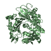
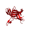
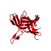
 Components Components | FIBER KNOB DOMAIN | ||||||
 Keywords Keywords | VIRAL PROTEIN / BETA-SANDWICH | ||||||
| Function / homology | : / Turkey Adenovirus 3 Fibre protein-like, head domain / Adenovirus Type 5 Fiber Protein (Receptor Binding Domain) - #50 / Adenovirus Type 5 Fiber Protein (Receptor Binding Domain) / Sandwich / Mainly Beta / PHOSPHATE ION / Fiber knob domain Function and homology information Function and homology information | ||||||
| Biological species |  TURKEY ADENOVIRUS 3 TURKEY ADENOVIRUS 3 | ||||||
| Method |  X-RAY DIFFRACTION / SYNCHROTRON / MOLECULAR REPLACEMENT / Resolution: 2.3 Å X-RAY DIFFRACTION / SYNCHROTRON / MOLECULAR REPLACEMENT / Resolution: 2.3 Å | ||||||
 Authors Authors | Singh, A.K. / van Raaij, M.J. | ||||||
 Citation Citation | Journal: PLoS ONE / Year: 2015 Title: Structure and Sialyllactose Binding of the Carboxy-Terminal Head Domain of the Fibre from a Siadenovirus, Turkey Adenovirus 3. Authors: Singh, A.K. / Berbis, M.A. / Ballmann, M.Z. / Kilcoyne, M. / Menendez, M. / Nguyen, T.H. / Joshi, L. / Canada, F.J. / Jimenez-Barbero, J. / Benko, M. / Harrach, B. / van Raaij, M.J. | ||||||
| History |
|
- Structure visualization
| Structure viewer | Molecule: MolmilJmol/JSmol |
|---|
- Downloads & links
-Download
| PDBx/mmCIF format | 4cw8.cif.gz | 44.1 KB | Display | PDBx/mmCIF format |
|---|---|---|---|---|
| PDB format | pdb4cw8.ent.gz | 30.1 KB | Display | PDB format |
| PDBx/mmJSON format | 4cw8.json.gz | Tree view | PDBx/mmJSON format | |
| Others |  Other downloads Other downloads |
-Validation report
| Arichive directory | https://data.pdbj.org/pub/pdb/validation_reports/cw/4cw8ftp://data.pdbj.org/pub/pdb/validation_reports/cw/4cw8 | HTTPS FTP |
|---|
-Related structure data
| Related structure data |  3zpeSC 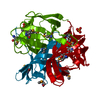 3zpfC  4d62C  4d63C S: Starting model for refinement C: citing same article ( |
|---|---|
| Similar structure data |


-Links
 PDBj
PDBj- Assembly
| Deposited unit | 
| ||||||||
|---|---|---|---|---|---|---|---|---|---|
| 1 | 
| ||||||||
| Unit cell |
| ||||||||
| Components on special symmetry positions |
|
-Components
| #1: Protein | Mass: 20835.156 Da / Num. of mol.: 1 / Fragment: HEAD DOMAIN, RESIDUES 12-165 / Mutation: YES Source method: isolated from a genetically manipulated source Source: (gene. exp.) TURKEY ADENOVIRUS 3 / Strain: AVIRULENT HEMORRHAGIC ENTERITIS VACCINE, LIVE VIRUS / Description: ARKO LABORATORIES, JEWELL IA / Variant: VETERINARY LICENSE NO. 337, SERIAL NO. 2081 / Production host:  | ||||
|---|---|---|---|---|---|
| #2: Chemical |   Mass: 94.971 Da / Num. of mol.: 2 / Source method: obtained synthetically / Formula: PO4 Mass: 94.971 Da / Num. of mol.: 2 / Source method: obtained synthetically / Formula: PO4#3: Water | ChemComp-HOH / |  Mass: 18.015 Da / Num. of mol.: 40 / Source method: isolated from a natural source / Formula: H2O Mass: 18.015 Da / Num. of mol.: 40 / Source method: isolated from a natural source / Formula: H2OSequence details | N-TERMINAL HIS-TAG, METHIONINE 354 WAS MUTATED TO ISOLEUCINE AND METHIONINE 376 WAS MUTATED TO ...N-TERMINAL HIS-TAG, METHIONINE | |
-Experimental details
-Experiment
| Experiment | Method: X-RAY DIFFRACTION / Number of used crystals: 1 |
|---|
- Sample preparation
| Crystal | Density Matthews: 2.1 Å3/Da / Density % sol: 42 % / Description: NONE |
|---|---|
| Crystal grow | pH: 7 Details: 10MM [2-(N-MORPHOLINO)ETHANESULFONIC ACID]-NAOH PH 6.0, 1M (NH4)2HPO4, 0.1 M IMIDAZOLE PH 7.0 |
-Data collection
| Diffraction | Mean temperature: 100 K |
|---|---|
| Diffraction source | Source: SYNCHROTRON / Site: ALBA  / Beamline: XALOC / Wavelength: 0.97946 / Beamline: XALOC / Wavelength: 0.97946 |
| Detector | Type: DECTRIS PILATUS 6M / Detector: PIXEL / Date: Jul 26, 2013 Details: PAIR OF KB MIRRORS FOR ADJUSTABLE FOCUSING, PLANE- ELLIPSOIDAL, THREE COATING STRIPES (SI, RH, IR) - IRELEC INSYNC |
| Radiation | Monochromator: CHANNEL-CUT DOUBLE CRYSTAL MONOCHROMATOR (CINEL), CRYOCOOLED, 6MM GAP Protocol: SINGLE WAVELENGTH / Monochromatic (M) / Laue (L): M / Scattering type: x-ray |
| Radiation wavelength | Wavelength: 0.97946 Å / Relative weight: 1 |
| Reflection | Resolution: 2.3→31.3 Å / Num. obs: 7327 / % possible obs: 100 % / Redundancy: 8.9 % / Biso Wilson estimate: 56.8 Å2 / Rmerge(I) obs: 0.06 / Net I/σ(I): 6.3 |
| Reflection shell | Resolution: 2.3→2.42 Å / Redundancy: 9.2 % / Rmerge(I) obs: 0.53 / Mean I/σ(I) obs: 1.4 / % possible all: 100 |
- Processing
| Software |
| ||||||||||||||||||||||||||||||||||||||||||||||||||||||||||||||||||||||||||||||||||||||||||||||||||||||||||||||||||||||||||||||||||||||||||||||||||||||||||||||||||||||||||||||||||||||
|---|---|---|---|---|---|---|---|---|---|---|---|---|---|---|---|---|---|---|---|---|---|---|---|---|---|---|---|---|---|---|---|---|---|---|---|---|---|---|---|---|---|---|---|---|---|---|---|---|---|---|---|---|---|---|---|---|---|---|---|---|---|---|---|---|---|---|---|---|---|---|---|---|---|---|---|---|---|---|---|---|---|---|---|---|---|---|---|---|---|---|---|---|---|---|---|---|---|---|---|---|---|---|---|---|---|---|---|---|---|---|---|---|---|---|---|---|---|---|---|---|---|---|---|---|---|---|---|---|---|---|---|---|---|---|---|---|---|---|---|---|---|---|---|---|---|---|---|---|---|---|---|---|---|---|---|---|---|---|---|---|---|---|---|---|---|---|---|---|---|---|---|---|---|---|---|---|---|---|---|---|---|---|---|
| Refinement | Method to determine structure: MOLECULAR REPLACEMENT Starting model: PDB ENTRY 3ZPE Resolution: 2.3→30 Å / Cor.coef. Fo:Fc: 0.964 / Cor.coef. Fo:Fc free: 0.934 / SU B: 9.997 / SU ML: 0.231 / Cross valid method: THROUGHOUT / ESU R: 0.318 / ESU R Free: 0.226 / Stereochemistry target values: MAXIMUM LIKELIHOOD Details: HYDROGENS HAVE BEEN ADDED IN THE RIDING POSITIONS. U VALUES REFINED INDIVIDUALLY
| ||||||||||||||||||||||||||||||||||||||||||||||||||||||||||||||||||||||||||||||||||||||||||||||||||||||||||||||||||||||||||||||||||||||||||||||||||||||||||||||||||||||||||||||||||||||
| Solvent computation | Ion probe radii: 0.8 Å / Shrinkage radii: 0.8 Å / VDW probe radii: 1.2 Å / Solvent model: MASK | ||||||||||||||||||||||||||||||||||||||||||||||||||||||||||||||||||||||||||||||||||||||||||||||||||||||||||||||||||||||||||||||||||||||||||||||||||||||||||||||||||||||||||||||||||||||
| Displacement parameters | Biso mean: 64.861 Å2
| ||||||||||||||||||||||||||||||||||||||||||||||||||||||||||||||||||||||||||||||||||||||||||||||||||||||||||||||||||||||||||||||||||||||||||||||||||||||||||||||||||||||||||||||||||||||
| Refinement step | Cycle: LAST / Resolution: 2.3→30 Å
| ||||||||||||||||||||||||||||||||||||||||||||||||||||||||||||||||||||||||||||||||||||||||||||||||||||||||||||||||||||||||||||||||||||||||||||||||||||||||||||||||||||||||||||||||||||||
| Refine LS restraints |
|