pullulanase activity / polysaccharide catabolic process / serine-type endopeptidase inhibitor activity / extracellular region / metal ion binding Similarity search - Function
Cereal seed allergen/trypsin and alpha-amylase inhibitor, conserved site / Cereal trypsin/alpha-amylase inhibitors family signature. / Cereal seed allergen/grain softness/trypsin and alpha-amylase inhibitor / Alpha-1,6-glucosidases, pullulanase-type / Alpha-1,6-glucosidases, pullulanase-type, C-terminal / Pullulanase, N2 domain / Alpha-1,6-glucosidases, pullulanase-type, C-terminal / Pullulanase N2 domain / Rab geranylgeranyltransferase alpha-subunit, insert domain / Plant lipid-transfer and hydrophobic proteins ...Cereal seed allergen/trypsin and alpha-amylase inhibitor, conserved site / Cereal trypsin/alpha-amylase inhibitors family signature. / Cereal seed allergen/grain softness/trypsin and alpha-amylase inhibitor / Alpha-1,6-glucosidases, pullulanase-type / Alpha-1,6-glucosidases, pullulanase-type, C-terminal / Pullulanase, N2 domain / Alpha-1,6-glucosidases, pullulanase-type, C-terminal / Pullulanase N2 domain / Rab geranylgeranyltransferase alpha-subunit, insert domain / Plant lipid-transfer and hydrophobic proteins / Hydrophobic Seed Protein / Protease inhibitor/seed storage/LTP family / Plant lipid transfer protein / seed storage protein / trypsin-alpha amylase inhibitor domain family / Bifunctional inhibitor/plant lipid transfer protein/seed storage helical domain / Bifunctional inhibitor/plant lipid transfer protein/seed storage helical domain superfamily / Glycoside hydrolase, family 13, N-terminal / Carbohydrate-binding module 48 (Isoamylase N-terminal domain) / Golgi alpha-mannosidase II / Glycosyl hydrolase, all-beta / Glycosidases / Immunoglobulin E-set / Glycoside hydrolase superfamily / TIM Barrel / Alpha-Beta Barrel / Immunoglobulin-like fold / Immunoglobulins / Immunoglobulin-like / Sandwich / Orthogonal Bundle / Mainly Beta / Mainly Alpha / Alpha Beta Similarity search - Domain/homology
SHEET DETERMINATION METHOD: DSSP THE SHEETS PRESENTED AS "AG" IN EACH CHAIN ON SHEET RECORDS BELOW ... SHEET DETERMINATION METHOD: DSSP THE SHEETS PRESENTED AS "AG" IN EACH CHAIN ON SHEET RECORDS BELOW IS ACTUALLY AN 8-STRANDED BARREL THIS IS REPRESENTED BY A 9-STRANDED SHEET IN WHICH THE FIRST AND LAST STRANDS ARE IDENTICAL. THE SHEETS PRESENTED AS "BF" IN EACH CHAIN ON SHEET RECORDS BELOW IS ACTUALLY AN 8-STRANDED BARREL THIS IS REPRESENTED BY A 9-STRANDED SHEET IN WHICH THE FIRST AND LAST STRANDS ARE IDENTICAL.
Mass: 18.015 Da / Num. of mol.: 98 / Source method: isolated from a natural source / Formula: H2O
Has protein modification
Y
Sequence details
THE VARIATIONS FROM THE ACCESSION NUMBER O48541 ARE AN ARG82LYS SUBSTITUTION AND A ...THE VARIATIONS FROM THE ACCESSION NUMBER O48541 ARE AN ARG82LYS SUBSTITUTION AND A NONE484VAL485THR486-TO- MET484ARG485ALA486 INSERTION-SUBSTITUTION. THE AMINO ACID CHANGES ARE LIKELY TO BE DEPENDENT ON BARLEY CULTIVAR 4 AMINO ACID DISCREPANCIES. THE SEQUENCE STRETCH BETWEEN RESIDUES 484-486 REFLECTS THAT THE CLONED CDNA IS FROM AN OFFSPRING OF THE UNP O48541 SOURCE AND THEREFORE A NATURAL VARIETY
-
Experimental details
-
Experiment
Experiment
Method: X-RAY DIFFRACTION / Number of used crystals: 1
-
Sample preparation
Crystal
Density Matthews: 2.5 Å3/Da / Density % sol: 50.83 % / Description: NONE
Crystal grow
Details: PROTEIN STOCK: PURIFIED 1:1 ENZYME:INHIBITOR COMPLEX (A280NM = 12.1) IN 50 MM MES PH 6.6, 250 MM NACL, 0.5 MM CACL2. RESERVOIR: 24% POLYETHYLENE GLYCOL (PEG) 8000 AND 0.05 M KH2PO4. 0.5 ...Details: PROTEIN STOCK: PURIFIED 1:1 ENZYME:INHIBITOR COMPLEX (A280NM = 12.1) IN 50 MM MES PH 6.6, 250 MM NACL, 0.5 MM CACL2. RESERVOIR: 24% POLYETHYLENE GLYCOL (PEG) 8000 AND 0.05 M KH2PO4. 0.5 MICROLITRE 0.1 M NAD WAS ADDED TO THE DROPLET TO A CONCENTRATION OF 0.01 M.
Resolution: 2.67→157.76 Å / Cor.coef. Fo:Fc: 0.891 / Cor.coef. Fo:Fc free: 0.847 / SU B: 12.208 / SU ML: 0.248 / Cross valid method: THROUGHOUT / ESU R Free: 0.088 / Stereochemistry target values: MAXIMUM LIKELIHOOD Details: HYDROGENS HAVE BEEN ADDED IN THE RIDING POSITIONS. U VALUES HAVE BEEN REFINED INDIVIDUALLY
Rfactor
Num. reflection
% reflection
Selection details
Rfree
0.293
3078
5.1 %
RANDOM
Rwork
0.255
-
-
-
obs
0.257
57661
96.3 %
-
Solvent computation
Ion probe radii: 0.8 Å / Shrinkage radii: 0.8 Å / VDW probe radii: 1.4 Å / Solvent model: BABINET MODEL WITH MASK
 Movie
Movie Controller

 Yorodumi
 Open data
 Basic information
 Components
 Keywords
 Function and homology information
 X-RAY DIFFRACTION /
  Authors
 Citation
 Structure visualization
 Downloads & links
 Other downloads
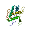
 PDBj
PDBj
 Assembly
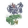

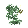
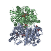
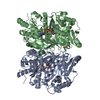
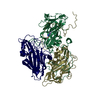
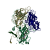
 KOMAGATAELLA PASTORIS (fungus) / Strain (production host): GS115 / References: UniProt: O48541, pullulanase
 Mass: 40.078 Da / Num. of mol.: 4 / Source method: obtained synthetically / Formula: Ca
 Mass: 18.015 Da / Num. of mol.: 98 / Source method: isolated from a natural source / Formula: H2O
 Sample preparation
 / Beamline: ID23-2 / Wavelength: 0.873
  Processing