 Movie
Movie Controller

[English] 日本語
 Yorodumi
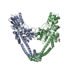
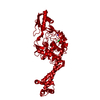
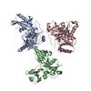
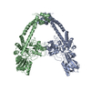
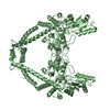
- PDB-4ckk: Apo structure of 55 kDa N-terminal domain of E. coli DNA gyrase A... -
+ Open data

- Basic information
| Entry | Database: PDB / ID: 4ckk | ||||||
|---|---|---|---|---|---|---|---|
| Title | Apo structure of 55 kDa N-terminal domain of E. coli DNA gyrase A subunit | ||||||
 Components Components | DNA GYRASE SUBUNIT A | ||||||
 Keywords Keywords | ISOMERASE / DNA GYRASE / TOPOISOMERASE / ANTIBIOTIC TARGET | ||||||
| Function / homology |  Function and homology information Function and homology informationDNA topoisomerase type II (double strand cut, ATP-hydrolyzing) complex / DNA negative supercoiling activity / DNA topoisomerase (ATP-hydrolysing) / DNA topological change / DNA-templated DNA replication / chromosome / response to antibiotic / DNA binding / ATP binding / cytoplasm Similarity search - Function | ||||||
| Biological species |  | ||||||
| Method |  X-RAY DIFFRACTION / SYNCHROTRON / MOLECULAR REPLACEMENT / Resolution: 1.9 Å X-RAY DIFFRACTION / SYNCHROTRON / MOLECULAR REPLACEMENT / Resolution: 1.9 Å | ||||||
 Authors Authors | Hearnshaw, S.J. / Edwards, M.J. / Stevenson, C.E.M. / Lawson, D.M. / Maxwell, A. | ||||||
 Citation Citation | Journal: J.Mol.Biol. / Year: 2014 Title: A New Crystal Structure of the Bifunctional Antibiotic Simocyclinone D8 Bound to DNA Gyrase Gives Fresh Insight Into the Mechanism of Inhibition. Authors: Hearnshaw, S.J. / Edwards, M.J. / Stevenson, C.E. / Lawson, D.M. / Maxwell, A. | ||||||
| History |
|
- Structure visualization
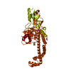
| Structure viewer | Molecule: MolmilJmol/JSmol |
|---|
- Downloads & links
-Download
| PDBx/mmCIF format | 4ckk.cif.gz | 761.3 KB | Display | PDBx/mmCIF format |
|---|---|---|---|---|
| PDB format | pdb4ckk.ent.gz | 633.7 KB | Display | PDB format |
| PDBx/mmJSON format | 4ckk.json.gz | Tree view | PDBx/mmJSON format | |
| Others |  Other downloads Other downloads |
-Validation report
| Arichive directory | https://data.pdbj.org/pub/pdb/validation_reports/ck/4ckkftp://data.pdbj.org/pub/pdb/validation_reports/ck/4ckk | HTTPS FTP |
|---|
-Related structure data
| Related structure data |  4cklC  2y3pS C: citing same article ( S: Starting model for refinement |
|---|---|
| Similar structure data |


-Links
 PDBj
PDBj

- Assembly
| Deposited unit | 
| ||||||||
|---|---|---|---|---|---|---|---|---|---|
| 1 | 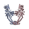
| ||||||||
| 2 | 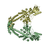
| ||||||||
| Unit cell |
|
-Components
| #1: Protein | Mass: 55381.398 Da / Num. of mol.: 4 / Fragment: 55 KDA N-TERMINAL DOMAIN, RESIDUES 30-522 Source method: isolated from a genetically manipulated source Source: (gene. exp.) References: UniProt: P0AES5, UniProt: A0A0H3JH39*PLUS, EC: 5.99.1.3 #2: Water | ChemComp-HOH / |  Mass: 18.015 Da / Num. of mol.: 1056 / Source method: isolated from a natural source / Formula: H2O Mass: 18.015 Da / Num. of mol.: 1056 / Source method: isolated from a natural source / Formula: H2O |
|---|
-Experimental details
-Experiment
| Experiment | Method: X-RAY DIFFRACTION / Number of used crystals: 1 |
|---|
- Sample preparation
| Crystal | Density Matthews: 2.96 Å3/Da / Density % sol: 59 % / Description: NONE |
|---|---|
| Crystal grow | pH: 8.5 / Details: pH 8.5 |
-Data collection
| Diffraction | Mean temperature: 100 K |
|---|---|
| Diffraction source | Source: SYNCHROTRON / Site: Diamond  / Beamline: I24 / Wavelength: 0.9686 / Beamline: I24 / Wavelength: 0.9686 |
| Detector | Type: DECTRIS PILATUS 6M / Detector: PIXEL / Date: May 14, 2012 |
| Radiation | Protocol: SINGLE WAVELENGTH / Monochromatic (M) / Laue (L): M / Scattering type: x-ray |
| Radiation wavelength | Wavelength: 0.9686 Å / Relative weight: 1 |
| Reflection | Resolution: 1.9→81.51 Å / Num. obs: 194509 / % possible obs: 97 % / Observed criterion σ(I): 0 / Redundancy: 2.9 % / Biso Wilson estimate: 23.6 Å2 / Rmerge(I) obs: 0.07 / Net I/σ(I): 11 |
| Reflection shell | Resolution: 1.9→1.95 Å / Redundancy: 2.6 % / Rmerge(I) obs: 0.49 / Mean I/σ(I) obs: 2.7 / % possible all: 94.7 |
- Processing
| Software |
| ||||||||||||||||||||||||||||||||||||||||||||||||||||||||||||||||||||||||||||||||||||||||||||||||||||||||||||||||||||||||||||||||||||||||||||||||||||||||||||||||||||||||||||||||||||||
|---|---|---|---|---|---|---|---|---|---|---|---|---|---|---|---|---|---|---|---|---|---|---|---|---|---|---|---|---|---|---|---|---|---|---|---|---|---|---|---|---|---|---|---|---|---|---|---|---|---|---|---|---|---|---|---|---|---|---|---|---|---|---|---|---|---|---|---|---|---|---|---|---|---|---|---|---|---|---|---|---|---|---|---|---|---|---|---|---|---|---|---|---|---|---|---|---|---|---|---|---|---|---|---|---|---|---|---|---|---|---|---|---|---|---|---|---|---|---|---|---|---|---|---|---|---|---|---|---|---|---|---|---|---|---|---|---|---|---|---|---|---|---|---|---|---|---|---|---|---|---|---|---|---|---|---|---|---|---|---|---|---|---|---|---|---|---|---|---|---|---|---|---|---|---|---|---|---|---|---|---|---|---|---|
| Refinement | Method to determine structure: MOLECULAR REPLACEMENT Starting model: PDB ENTRY 2Y3P Resolution: 1.9→81.51 Å / Cor.coef. Fo:Fc: 0.961 / Cor.coef. Fo:Fc free: 0.956 / SU B: 4.872 / SU ML: 0.072 / Cross valid method: THROUGHOUT / ESU R: 0.121 / ESU R Free: 0.109 / Stereochemistry target values: MAXIMUM LIKELIHOOD Details: HYDROGENS HAVE BEEN ADDED IN THE RIDING POSITIONS. U VALUES WITH TLS ADDED
| ||||||||||||||||||||||||||||||||||||||||||||||||||||||||||||||||||||||||||||||||||||||||||||||||||||||||||||||||||||||||||||||||||||||||||||||||||||||||||||||||||||||||||||||||||||||
| Solvent computation | Ion probe radii: 0.8 Å / Shrinkage radii: 0.8 Å / VDW probe radii: 1.2 Å / Solvent model: MASK | ||||||||||||||||||||||||||||||||||||||||||||||||||||||||||||||||||||||||||||||||||||||||||||||||||||||||||||||||||||||||||||||||||||||||||||||||||||||||||||||||||||||||||||||||||||||
| Displacement parameters | Biso mean: 32.3 Å2
| ||||||||||||||||||||||||||||||||||||||||||||||||||||||||||||||||||||||||||||||||||||||||||||||||||||||||||||||||||||||||||||||||||||||||||||||||||||||||||||||||||||||||||||||||||||||
| Refinement step | Cycle: LAST / Resolution: 1.9→81.51 Å
| ||||||||||||||||||||||||||||||||||||||||||||||||||||||||||||||||||||||||||||||||||||||||||||||||||||||||||||||||||||||||||||||||||||||||||||||||||||||||||||||||||||||||||||||||||||||
| Refine LS restraints |
|