 Movie
Movie Controller

[English] 日本語
 Yorodumi
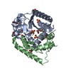
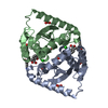
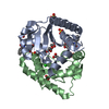
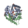
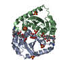
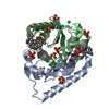
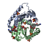
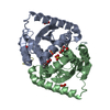
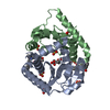
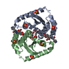
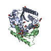
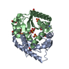
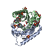
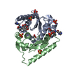
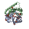
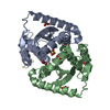
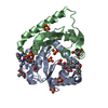
- PDB-4cf2: Interrogating HIV integrase for compounds that bind- a SAMPL challenge -
+ Open data

- Basic information
| Entry | Database: PDB / ID: 4cf2 | ||||||
|---|---|---|---|---|---|---|---|
| Title | Interrogating HIV integrase for compounds that bind- a SAMPL challenge | ||||||
 Components Components | INTEGRASE | ||||||
 Keywords Keywords | TRANSFERASE / HIV INTEGRASE / STRUCTURE BASED DRUG DESIGN | ||||||
| Function / homology |  Function and homology information Function and homology informationHIV-1 retropepsin / symbiont-mediated activation of host apoptosis / retroviral ribonuclease H / exoribonuclease H / exoribonuclease H activity / DNA integration / viral genome integration into host DNA / establishment of integrated proviral latency / RNA-directed DNA polymerase / RNA stem-loop binding ...HIV-1 retropepsin / symbiont-mediated activation of host apoptosis / retroviral ribonuclease H / exoribonuclease H / exoribonuclease H activity / DNA integration / viral genome integration into host DNA / establishment of integrated proviral latency / RNA-directed DNA polymerase / RNA stem-loop binding / viral penetration into host nucleus / host multivesicular body / RNA-directed DNA polymerase activity / RNA-DNA hybrid ribonuclease activity / Transferases; Transferring phosphorus-containing groups; Nucleotidyltransferases / host cell / viral nucleocapsid / endonuclease activity / DNA recombination / DNA-directed DNA polymerase / aspartic-type endopeptidase activity / Hydrolases; Acting on ester bonds / host cell cytoplasm / DNA-directed DNA polymerase activity / symbiont-mediated suppression of host gene expression / viral translational frameshifting / symbiont entry into host cell / lipid binding / host cell nucleus / host cell plasma membrane / virion membrane / structural molecule activity / proteolysis / DNA binding / zinc ion binding Similarity search - Function | ||||||
| Biological species |   HUMAN IMMUNODEFICIENCY VIRUS 1 HUMAN IMMUNODEFICIENCY VIRUS 1 | ||||||
| Method |  X-RAY DIFFRACTION / SYNCHROTRON / MOLECULAR REPLACEMENT / Resolution: 1.95 Å X-RAY DIFFRACTION / SYNCHROTRON / MOLECULAR REPLACEMENT / Resolution: 1.95 Å | ||||||
 Authors Authors | Peat, T.S. | ||||||
 Citation Citation | Journal: J.Comput.Aided Mol.Des. / Year: 2014 Title: Interrogating HIV Integrase for Compounds that Bind- a Sampl Challenge. Authors: Peat, T.S. / Dolezal, O. / Newman, J. / Mobley, D. / Deadman, J.J. | ||||||
| History |
|
- Structure visualization
| Structure viewer | Molecule: MolmilJmol/JSmol |
|---|
- Downloads & links
-Download
| PDBx/mmCIF format | 4cf2.cif.gz | 79 KB | Display | PDBx/mmCIF format |
|---|---|---|---|---|
| PDB format | pdb4cf2.ent.gz | 58.8 KB | Display | PDB format |
| PDBx/mmJSON format | 4cf2.json.gz | Tree view | PDBx/mmJSON format | |
| Others |  Other downloads Other downloads |
-Validation report
| Arichive directory | https://data.pdbj.org/pub/pdb/validation_reports/cf/4cf2ftp://data.pdbj.org/pub/pdb/validation_reports/cf/4cf2 | HTTPS FTP |
|---|
-Related structure data
| Related structure data |  4ce9C  4ceaC  4cebC 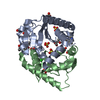 4cecC  4cedC 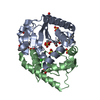 4ceeC  4cefC 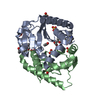 4ceoC  4ceqC 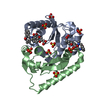 4cerC 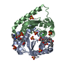 4cesC 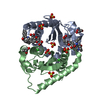 4cezC  4cf0C 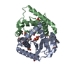 4cf1C 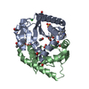 4cf8C 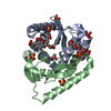 4cf9C 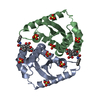 4cfaC 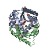 4cfbC 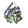 4cfcC  4cfdC 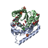 4cgdC 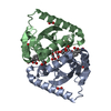 4cgfC  4cggC  4cghC 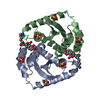 4cgiC 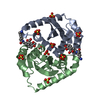 4cgjC  4chnC 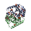 4choC  4chpC  4chqC  4chyC  4chzC  4cieC  4cifC  4cigC  4cj3C  4cj4C  4cj5C  4cjeC  4cjfC  4cjkC 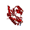 4cjlC 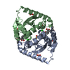 4cjpC  4cjqC  4cjrC  4cjsC  4cjtC  4cjuC  4cjvC  4cjwC  4ck1C 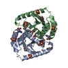 4ck2C  4ck3C  4ovlC 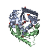 3zsqS C: citing same article ( S: Starting model for refinement |
|---|---|
| Similar structure data |


-Links
 PDBj
PDBj
- Assembly
| Deposited unit | 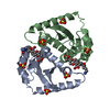
| ||||||||
|---|---|---|---|---|---|---|---|---|---|
| 1 |
| ||||||||
| Unit cell |
|
-Components
| #1: Protein | Mass: 20044.672 Da / Num. of mol.: 2 / Fragment: CATALYTIC DOMAIN, RESIDUES 50-212 / Mutation: YES Source method: isolated from a genetically manipulated source Source: (gene. exp.) HUMAN IMMUNODEFICIENCY VIRUS 1 / Strain: TYPE1 / Production host:  References: UniProt: Q76353, UniProt: P12497*PLUS, DNA-directed DNA polymerase #2: Chemical | ChemComp-SO4 /   Mass: 96.063 Da / Num. of mol.: 6 / Source method: obtained synthetically / Formula: SO4 Mass: 96.063 Da / Num. of mol.: 6 / Source method: obtained synthetically / Formula: SO4#3: Chemical |   Mass: 482.526 Da / Num. of mol.: 2 / Source method: obtained synthetically / Formula: C26H30N2O7 Mass: 482.526 Da / Num. of mol.: 2 / Source method: obtained synthetically / Formula: C26H30N2O7#4: Water | ChemComp-HOH / |  Mass: 18.015 Da / Num. of mol.: 75 / Source method: isolated from a natural source / Formula: H2O Mass: 18.015 Da / Num. of mol.: 75 / Source method: isolated from a natural source / Formula: H2O |
|---|
-Experimental details
-Experiment
| Experiment | Method: X-RAY DIFFRACTION / Number of used crystals: 1 |
|---|
- Sample preparation
| Crystal | Density Matthews: 2.95 Å3/Da / Density % sol: 58 % / Description: NONE |
|---|---|
| Crystal grow | pH: 5.5 Details: THE PROTEIN WAS CONCENTRATED TO 5.5 MG/ML IN 40 MM TRIS PH 8.0, 250 MM NACL, 30 MM MGCL2, 5 MM DTT AND SET UP IN A 1:1 RATIO WITH 1.6 TO 2.0 M AMMONIUM SULFATE, 100 MM SODIUM ACETATE BUFFER PH 5.0 TO 5.5. |
-Data collection
| Diffraction | Mean temperature: 100 K |
|---|---|
| Diffraction source | Source: SYNCHROTRON / Site: Australian Synchrotron  / Beamline: MX1 / Wavelength: 0.95367 / Beamline: MX1 / Wavelength: 0.95367 |
| Detector | Type: ADSC CCD / Detector: CCD / Date: Aug 22, 2009 |
| Radiation | Protocol: SINGLE WAVELENGTH / Monochromatic (M) / Laue (L): M / Scattering type: x-ray |
| Radiation wavelength | Wavelength: 0.95367 Å / Relative weight: 1 |
| Reflection | Resolution: 1.95→45.2 Å / Num. obs: 27514 / % possible obs: 99.9 % / Observed criterion σ(I): 1 / Redundancy: 5.5 % / Rmerge(I) obs: 0.07 / Net I/σ(I): 18.3 |
| Reflection shell | Resolution: 1.95→2.06 Å / Redundancy: 5.4 % / Rmerge(I) obs: 0.53 / Mean I/σ(I) obs: 2.9 / % possible all: 100 |
- Processing
| Software |
| ||||||||||||||||||||||||||||||||||||||||||||||||||||||||||||||||||||||||||||||||||||||||||||||||||||||||||||||||||||||||||||||||||||||||||||||||||||||||||||||||||||||||||||||||||||||
|---|---|---|---|---|---|---|---|---|---|---|---|---|---|---|---|---|---|---|---|---|---|---|---|---|---|---|---|---|---|---|---|---|---|---|---|---|---|---|---|---|---|---|---|---|---|---|---|---|---|---|---|---|---|---|---|---|---|---|---|---|---|---|---|---|---|---|---|---|---|---|---|---|---|---|---|---|---|---|---|---|---|---|---|---|---|---|---|---|---|---|---|---|---|---|---|---|---|---|---|---|---|---|---|---|---|---|---|---|---|---|---|---|---|---|---|---|---|---|---|---|---|---|---|---|---|---|---|---|---|---|---|---|---|---|---|---|---|---|---|---|---|---|---|---|---|---|---|---|---|---|---|---|---|---|---|---|---|---|---|---|---|---|---|---|---|---|---|---|---|---|---|---|---|---|---|---|---|---|---|---|---|---|---|
| Refinement | Method to determine structure: MOLECULAR REPLACEMENT Starting model: PDB ENTRY 3ZSQ Resolution: 1.95→31.42 Å / Cor.coef. Fo:Fc: 0.945 / Cor.coef. Fo:Fc free: 0.935 / SU B: 3.508 / SU ML: 0.101 / Cross valid method: THROUGHOUT / ESU R: 0.166 / ESU R Free: 0.142 / Stereochemistry target values: MAXIMUM LIKELIHOOD Details: HYDROGENS HAVE BEEN ADDED IN THE RIDING POSITIONS. U VALUES REFINED INDIVIDUALLY
| ||||||||||||||||||||||||||||||||||||||||||||||||||||||||||||||||||||||||||||||||||||||||||||||||||||||||||||||||||||||||||||||||||||||||||||||||||||||||||||||||||||||||||||||||||||||
| Solvent computation | Ion probe radii: 0.8 Å / Shrinkage radii: 0.8 Å / VDW probe radii: 1.2 Å / Solvent model: MASK | ||||||||||||||||||||||||||||||||||||||||||||||||||||||||||||||||||||||||||||||||||||||||||||||||||||||||||||||||||||||||||||||||||||||||||||||||||||||||||||||||||||||||||||||||||||||
| Displacement parameters | Biso mean: 32.755 Å2
| ||||||||||||||||||||||||||||||||||||||||||||||||||||||||||||||||||||||||||||||||||||||||||||||||||||||||||||||||||||||||||||||||||||||||||||||||||||||||||||||||||||||||||||||||||||||
| Refinement step | Cycle: LAST / Resolution: 1.95→31.42 Å
| ||||||||||||||||||||||||||||||||||||||||||||||||||||||||||||||||||||||||||||||||||||||||||||||||||||||||||||||||||||||||||||||||||||||||||||||||||||||||||||||||||||||||||||||||||||||
| Refine LS restraints |
|