 Movie
Movie Controller


+ Open data

- Basic information
| Entry | Database: PDB / ID: 4c82 | ||||||
|---|---|---|---|---|---|---|---|
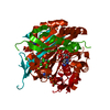
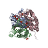
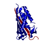
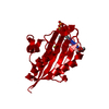
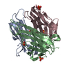
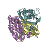
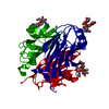
| Title | IspF (Plasmodium falciparum) unliganded structure | ||||||
 Components Components | 2-C-METHYL-D-ERYTHRITOL 2,4-CYCLODIPHOSPHATE SYNTHASE | ||||||
 Keywords Keywords | LYASE | ||||||
| Function / homology |  Function and homology information Function and homology information2-C-methyl-D-erythritol 2,4-cyclodiphosphate synthase / 2-C-methyl-D-erythritol 2,4-cyclodiphosphate synthase activity / apicoplast / isopentenyl diphosphate biosynthetic process, methylerythritol 4-phosphate pathway / terpenoid biosynthetic process / metal ion binding Similarity search - Function | ||||||
| Biological species |  | ||||||
| Method |  X-RAY DIFFRACTION / MOLECULAR REPLACEMENT / Resolution: 2 Å X-RAY DIFFRACTION / MOLECULAR REPLACEMENT / Resolution: 2 Å | ||||||
 Authors Authors | O'Rourke, P.E.F. / Kalinowska-Tluscik, J. / Fyfe, P.K. / Dawson, A. / Hunter, W.N. | ||||||
 Citation Citation | Journal: Bmc Struct.Biol. / Year: 2014 Title: Crystal Structures of Ispf from Plasmodium Falciparum and Burkholderia Cenocepacia: Comparisons Inform Antimicrobial Drug Target Assessment. Authors: O Rourke, P.E. / Kalinowska-Tluscik, J. / Fyfe, P.K. / Dawson, A. / Hunter, W.N. | ||||||
| History |
|
- Structure visualization
| Structure viewer | Molecule: MolmilJmol/JSmol |
|---|
- Downloads & links
-Download
| PDBx/mmCIF format | 4c82.cif.gz | 48.9 KB | Display | PDBx/mmCIF format |
|---|---|---|---|---|
| PDB format | pdb4c82.ent.gz | 33.4 KB | Display | PDB format |
| PDBx/mmJSON format | 4c82.json.gz | Tree view | PDBx/mmJSON format | |
| Others |  Other downloads Other downloads |
-Validation report
| Arichive directory | https://data.pdbj.org/pub/pdb/validation_reports/c8/4c82ftp://data.pdbj.org/pub/pdb/validation_reports/c8/4c82 | HTTPS FTP |
|---|
-Related structure data
| Related structure data |  4c81SC  4c8eC  4c8gC  4c8iC S: Starting model for refinement C: citing same article ( |
|---|---|
| Similar structure data |

-Links
 PDBj
PDBj- Assembly
| Deposited unit | 
| |||||||||
|---|---|---|---|---|---|---|---|---|---|---|
| 1 | 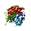
| |||||||||
| Unit cell |
| |||||||||
| Components on special symmetry positions |
|
-Components
| #1: Protein | Mass: 20574.588 Da / Num. of mol.: 1 Fragment: MATURE PROTEIN (APICOPLAST-TARGETING SEQUENCE OMITTED), RESIDUES 60-240 Mutation: YES Source method: isolated from a genetically manipulated source Source: (gene. exp.) Strain: 3D7 / Plasmid: PET15BTEV / Production host:  References: UniProt: P62368, 2-C-methyl-D-erythritol 2,4-cyclodiphosphate synthase | ||||||
|---|---|---|---|---|---|---|---|
| #2: Chemical |   Mass: 65.409 Da / Num. of mol.: 2 / Source method: obtained synthetically / Formula: Zn Mass: 65.409 Da / Num. of mol.: 2 / Source method: obtained synthetically / Formula: Zn#3: Chemical | ChemComp-SO4 / |   Mass: 96.063 Da / Num. of mol.: 1 / Source method: obtained synthetically / Formula: SO4 Mass: 96.063 Da / Num. of mol.: 1 / Source method: obtained synthetically / Formula: SO4#4: Water | ChemComp-HOH / |  Mass: 18.015 Da / Num. of mol.: 94 / Source method: isolated from a natural source / Formula: H2O Mass: 18.015 Da / Num. of mol.: 94 / Source method: isolated from a natural source / Formula: H2OSequence details | RESIDUES 1-59, A PREDICTED APICOPLAST TARGETING SEQUENCE, HAVE BEEN OMITTED FROM THE CONSTRUCT USED ...RESIDUES 1-59, A PREDICTED APICOPLAST | |
-Experimental details
-Experiment
| Experiment | Method: X-RAY DIFFRACTION / Number of used crystals: 1 |
|---|
- Sample preparation
| Crystal | Density Matthews: 3.4 Å3/Da / Density % sol: 64 % / Description: NONE |
|---|---|
| Crystal grow | Temperature: 293 K / Method: vapor diffusion, hanging drop / pH: 5.5 Details: PROTEIN WAS CRYSTALLISED BY HANGING-DROP VAPOUR DIFFUSION. PFISPF SAMPLE IN 100 MM KCL, 100 MM L-ARGININE, 2 MM MGCL2, 50 MM CHES, PH 9.5; WAS CONCENTRATED TO 6 MG/ML, AND HAD FOSMIDOMYCIN ...Details: PROTEIN WAS CRYSTALLISED BY HANGING-DROP VAPOUR DIFFUSION. PFISPF SAMPLE IN 100 MM KCL, 100 MM L-ARGININE, 2 MM MGCL2, 50 MM CHES, PH 9.5; WAS CONCENTRATED TO 6 MG/ML, AND HAD FOSMIDOMYCIN ADDED (20 MM FINAL CONCENTRATION). CRYSTALS WERE OBTAINED BY MIXING 2 MICROLITES OF PROTEIN WITH 2 MICROLITRES OF RESERVOIR SOLUTION AT 20 DEGREES C. RESERVOIR SOLUTION: 1.8-2.5 M (NH4)2SO4, 5MM ZNCL2, 100 MM BIS-TRIS, PH 5.5. SATURATED SUCROSE WAS USED AS A CRYO-PROTECTANT DURING COLLECTION. |
-Data collection
| Diffraction | Mean temperature: 100 K |
|---|---|
| Diffraction source | Source: ROTATING ANODE / Type: RIGAKU MICROMAX-007 HF / Wavelength: 1.5418 |
| Detector | Type: RIGAKU RAXIS IV PLUS PLUS / Detector: IMAGE PLATE / Date: Oct 12, 2010 |
| Radiation | Protocol: SINGLE WAVELENGTH / Monochromatic (M) / Laue (L): M / Scattering type: x-ray |
| Radiation wavelength | Wavelength: 1.5418 Å / Relative weight: 1 |
| Reflection | Resolution: 2→29.71 Å / Num. obs: 18068 / % possible obs: 98.6 % / Observed criterion σ(I): 2 / Redundancy: 2.3 % / Biso Wilson estimate: 32 Å2 / Rmerge(I) obs: 0.09 / Net I/σ(I): 5.3 |
| Reflection shell | Resolution: 2→2.11 Å / Redundancy: 2.2 % / Rmerge(I) obs: 0.48 / Mean I/σ(I) obs: 2 / % possible all: 97 |
- Processing
| Software |
| ||||||||||||||||||||||||||||||||||||||||||||||||||||||||||||||||||||||||||||||||||||||||||||||||||||||||||||||||||||||||||||||||||||||||||||||||||||||||||||||||||||||||||||||||||||||
|---|---|---|---|---|---|---|---|---|---|---|---|---|---|---|---|---|---|---|---|---|---|---|---|---|---|---|---|---|---|---|---|---|---|---|---|---|---|---|---|---|---|---|---|---|---|---|---|---|---|---|---|---|---|---|---|---|---|---|---|---|---|---|---|---|---|---|---|---|---|---|---|---|---|---|---|---|---|---|---|---|---|---|---|---|---|---|---|---|---|---|---|---|---|---|---|---|---|---|---|---|---|---|---|---|---|---|---|---|---|---|---|---|---|---|---|---|---|---|---|---|---|---|---|---|---|---|---|---|---|---|---|---|---|---|---|---|---|---|---|---|---|---|---|---|---|---|---|---|---|---|---|---|---|---|---|---|---|---|---|---|---|---|---|---|---|---|---|---|---|---|---|---|---|---|---|---|---|---|---|---|---|---|---|
| Refinement | Method to determine structure: MOLECULAR REPLACEMENT Starting model: PDB ENTRY 4C81 Resolution: 2→29.72 Å / Cor.coef. Fo:Fc: 0.944 / Cor.coef. Fo:Fc free: 0.94 / SU B: 3.406 / SU ML: 0.096 / Cross valid method: THROUGHOUT / ESU R: 0.133 / ESU R Free: 0.129 / Stereochemistry target values: MAXIMUM LIKELIHOOD Details: HYDROGENS HAVE BEEN ADDED IN THE RIDING POSITIONS. RESIDUES 82-97 AND 144-151 ARE DISORDERED. HYDROGENS HAVE BEEN USED IF PRESENT IN THE INPUT U VALUES REFINED INDIVIDUALLY
| ||||||||||||||||||||||||||||||||||||||||||||||||||||||||||||||||||||||||||||||||||||||||||||||||||||||||||||||||||||||||||||||||||||||||||||||||||||||||||||||||||||||||||||||||||||||
| Solvent computation | Ion probe radii: 0.8 Å / Shrinkage radii: 0.8 Å / VDW probe radii: 1.2 Å / Solvent model: MASK | ||||||||||||||||||||||||||||||||||||||||||||||||||||||||||||||||||||||||||||||||||||||||||||||||||||||||||||||||||||||||||||||||||||||||||||||||||||||||||||||||||||||||||||||||||||||
| Displacement parameters | Biso mean: 37.598 Å2
| ||||||||||||||||||||||||||||||||||||||||||||||||||||||||||||||||||||||||||||||||||||||||||||||||||||||||||||||||||||||||||||||||||||||||||||||||||||||||||||||||||||||||||||||||||||||
| Refinement step | Cycle: LAST / Resolution: 2→29.72 Å
| ||||||||||||||||||||||||||||||||||||||||||||||||||||||||||||||||||||||||||||||||||||||||||||||||||||||||||||||||||||||||||||||||||||||||||||||||||||||||||||||||||||||||||||||||||||||
| Refine LS restraints |
|