 Movie
Movie Controller

[English] 日本語
 Yorodumi
- PDB-4c0x: The crystal strucuture of PpAzoR in complex with anthraquinone-2-... -
+ Open data

- Basic information
| Entry | Database: PDB / ID: 4c0x | ||||||
|---|---|---|---|---|---|---|---|
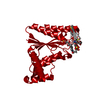
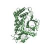
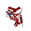
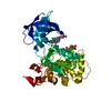
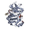
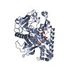
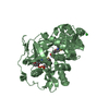
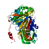
| Title | The crystal strucuture of PpAzoR in complex with anthraquinone-2- sulfonate | ||||||
 Components Components | FMN-DEPENDENT NADH-AZOREDUCTASE 1 | ||||||
 Keywords Keywords | OXIDOREDUCTASE / AZOREDUCTASE / NAD(P)H QUINONE OXIDOREDUCTASE | ||||||
| Function / homology |  Function and homology information Function and homology informationFMN-dependent NADH-azoreductase / oxidoreductase activity, acting on NAD(P)H as acceptor / Oxidoreductases; Acting on NADH or NADPH; With a quinone or similar compound as acceptor / oxidoreductase activity, acting on NAD(P)H, quinone or similar compound as acceptor / FMN binding / electron transfer activity Similarity search - Function | ||||||
| Biological species |  PSEUDOMONAS PUTIDA (bacteria) PSEUDOMONAS PUTIDA (bacteria) | ||||||
| Method |  X-RAY DIFFRACTION / SYNCHROTRON / MOLECULAR REPLACEMENT / Resolution: 1.499 Å X-RAY DIFFRACTION / SYNCHROTRON / MOLECULAR REPLACEMENT / Resolution: 1.499 Å | ||||||
 Authors Authors | Goncalves, A.M.D. / de Sanctis, D. / Bento, I. | ||||||
 Citation Citation | Journal: FEBS J. / Year: 2013 Title: The Crystal Structure of Pseudomonas Putida Azor: The Active Site Revisited. Authors: Goncalves, A.M.D. / Mendes, S. / De Sanctis, D. / Martins, L.O. / Bento, I. | ||||||
| History |
|
- Structure visualization
| Structure viewer | Molecule: MolmilJmol/JSmol |
|---|
- Downloads & links
-Download
| PDBx/mmCIF format | 4c0x.cif.gz | 62.8 KB | Display | PDBx/mmCIF format |
|---|---|---|---|---|
| PDB format | pdb4c0x.ent.gz | 44.3 KB | Display | PDB format |
| PDBx/mmJSON format | 4c0x.json.gz | Tree view | PDBx/mmJSON format | |
| Others |  Other downloads Other downloads |
-Validation report
| Arichive directory | https://data.pdbj.org/pub/pdb/validation_reports/c0/4c0xftp://data.pdbj.org/pub/pdb/validation_reports/c0/4c0x | HTTPS FTP |
|---|
-Related structure data
| Related structure data |  4c0wSC  4c14C S: Starting model for refinement C: citing same article ( |
|---|---|
| Similar structure data |

-Links
 PDBj
PDBj- Assembly
| Deposited unit | 
| ||||||||||||
|---|---|---|---|---|---|---|---|---|---|---|---|---|---|
| 1 | 
| ||||||||||||
| Unit cell |
| ||||||||||||
| Components on special symmetry positions |
|
-Components
-Protein , 1 types, 1 molecules A
| #1: Protein | Mass: 21414.156 Da / Num. of mol.: 1 Source method: isolated from a genetically manipulated source Details: FMN-BINDING PROTEIN, COMPLEXED TO ANTHRAQUINONE-2-SULFONATE Source: (gene. exp.) PSEUDOMONAS PUTIDA (bacteria) / Strain: MET94 / Production host: References: UniProt: Q88IY3, Oxidoreductases; Acting on other nitrogenous compounds as donors, NAD(P)H dehydrogenase (quinone) |
|---|
-Non-polymers , 5 types, 261 molecules 


| #2: Chemical | ChemComp-FMN /  Mass: 456.344 Da / Num. of mol.: 1 / Source method: obtained synthetically / Formula: C17H21N4O9P Mass: 456.344 Da / Num. of mol.: 1 / Source method: obtained synthetically / Formula: C17H21N4O9P | ||||
|---|---|---|---|---|---|
| #3: Chemical | ChemComp-AQN /  Mass: 288.275 Da / Num. of mol.: 1 / Source method: obtained synthetically / Formula: C14H8O5S Mass: 288.275 Da / Num. of mol.: 1 / Source method: obtained synthetically / Formula: C14H8O5S | ||||
| #4: Chemical |  Mass: 92.094 Da / Num. of mol.: 2 / Source method: obtained synthetically / Formula: C3H8O3 Mass: 92.094 Da / Num. of mol.: 2 / Source method: obtained synthetically / Formula: C3H8O3#5: Chemical |  Mass: 194.226 Da / Num. of mol.: 2 / Source method: obtained synthetically / Formula: C8H18O5 / Comment: precipitant*YM Mass: 194.226 Da / Num. of mol.: 2 / Source method: obtained synthetically / Formula: C8H18O5 / Comment: precipitant*YM#6: Water | ChemComp-HOH / | Mass: 18.015 Da / Num. of mol.: 255 / Source method: isolated from a natural source / Formula: H2O |
-Details
| Sequence details | THE GENOME OF PSEUDOMONAS PUTIDA STRAIN MET94 HAS NOT BEEN SEQUENCED TO DATE. THE HIGHEST HOMOLOGY ...THE GENOME OF PSEUDOMONA |
|---|
-Experimental details
-Experiment
| Experiment | Method: X-RAY DIFFRACTION / Number of used crystals: 1 |
|---|
- Sample preparation
| Crystal | Density Matthews: 2.8 Å3/Da / Density % sol: 56 % / Description: NONE |
|---|---|
| Crystal grow | Details: 1.8 M AMMONIUM SULFATE, 0.1 M HEPES, PH 7.0, 4% (V:V) PEG400 |
-Data collection
| Diffraction | Mean temperature: 100 K |
|---|---|
| Diffraction source | Source: SYNCHROTRON / Site: ESRF  / Beamline: ID14-1 / Wavelength: 0.9334 / Beamline: ID14-1 / Wavelength: 0.9334 |
| Detector | Type: ADSC QUANTUM 210 / Detector: CCD / Date: Nov 7, 2012 |
| Radiation | Protocol: SINGLE WAVELENGTH / Monochromatic (M) / Laue (L): M / Scattering type: x-ray |
| Radiation wavelength | Wavelength: 0.9334 Å / Relative weight: 1 |
| Reflection | Resolution: 1.5→47.5 Å / Num. obs: 40377 / % possible obs: 99.4 % / Observed criterion σ(I): 2 / Redundancy: 7.7 % / Biso Wilson estimate: 18.91 Å2 / Rmerge(I) obs: 0.06 / Net I/σ(I): 8.5 |
| Reflection shell | Resolution: 1.5→1.58 Å / Redundancy: 3.7 % / Rmerge(I) obs: 0.29 / Mean I/σ(I) obs: 2.7 / % possible all: 99.9 |
- Processing
| Software |
| |||||||||||||||||||||||||||||||||||||||||||||||||||||||||||||||||||||||||||||
|---|---|---|---|---|---|---|---|---|---|---|---|---|---|---|---|---|---|---|---|---|---|---|---|---|---|---|---|---|---|---|---|---|---|---|---|---|---|---|---|---|---|---|---|---|---|---|---|---|---|---|---|---|---|---|---|---|---|---|---|---|---|---|---|---|---|---|---|---|---|---|---|---|---|---|---|---|---|---|
| Refinement | Method to determine structure: MOLECULAR REPLACEMENT Starting model: PDB ENTRY 4C0W Resolution: 1.499→47.474 Å / SU ML: 0.14 / σ(F): 1.35 / Phase error: 19.85 / Stereochemistry target values: ML / Details: RESIDUES 201-203 ARE DISORDERED.
| |||||||||||||||||||||||||||||||||||||||||||||||||||||||||||||||||||||||||||||
| Solvent computation | Shrinkage radii: 0.9 Å / VDW probe radii: 1.11 Å / Solvent model: FLAT BULK SOLVENT MODEL | |||||||||||||||||||||||||||||||||||||||||||||||||||||||||||||||||||||||||||||
| Displacement parameters | Biso mean: 27.3 Å2 | |||||||||||||||||||||||||||||||||||||||||||||||||||||||||||||||||||||||||||||
| Refinement step | Cycle: LAST / Resolution: 1.499→47.474 Å
| |||||||||||||||||||||||||||||||||||||||||||||||||||||||||||||||||||||||||||||
| Refine LS restraints |
| |||||||||||||||||||||||||||||||||||||||||||||||||||||||||||||||||||||||||||||
| LS refinement shell |
|
