Protocol: SINGLE WAVELENGTH / Monochromatic (M) / Laue (L): M / Scattering type: x-ray
Radiation wavelength
Wavelength: 0.9795 Å / Relative weight: 1
Reflection
Resolution: 2.15→96.04 Å / Num. obs: 56465 / % possible obs: 98.3 % / Observed criterion σ(I): 2 / Redundancy: 4 % / Rmerge(I) obs: 0.05 / Net I/σ(I): 25.1
Reflection shell
Resolution: 2.15→2.21 Å / Redundancy: 2.7 % / Rmerge(I) obs: 0.65 / Mean I/σ(I) obs: 2.6 / % possible all: 97.3
-
Processing
Software
Name
Version
Classification
REFMAC
5.6.0111
refinement
xia2
datareduction
xia2
datascaling
SHELX
phasing
Refinement
Method to determine structure: SAD Starting model: NONE Resolution: 2.15→96.04 Å / Cor.coef. Fo:Fc: 0.956 / Cor.coef. Fo:Fc free: 0.947 / SU B: 11.366 / SU ML: 0.145 / Cross valid method: THROUGHOUT / ESU R: 0.2 / ESU R Free: 0.172 / Stereochemistry target values: MAXIMUM LIKELIHOOD Details: HYDROGENS HAVE BEEN ADDED IN THE RIDING POSITIONS. HYDROGENS HAVE BEEN USED IF PRESENT IN THE INPUT. U VALUES WITH TLS ADDED. DISORDERED REGIONS WERE MODELED STEREOCHEMICALLY
Rfactor
Num. reflection
% reflection
Selection details
Rfree
0.24427
2865
5.1 %
RANDOM
Rwork
0.2183
-
-
-
obs
0.21963
53600
98.28 %
-
Solvent computation
Ion probe radii: 0.8 Å / Shrinkage radii: 0.8 Å / VDW probe radii: 1.2 Å / Solvent model: BABINET MODEL WITH MASK
 Movie
Movie Controller

 Yorodumi
 Open data
 Basic information
 Components
 Keywords
 Function and homology information
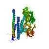
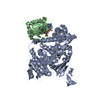
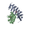
 STREPTOMYCES PHAGE PHIC31 (virus)
 X-RAY DIFFRACTION /
  Authors
 Citation
 Structure visualization
 Downloads & links
 Other downloads
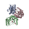


 PDBj
PDBj Assembly

 Mass: 18.015 Da / Num. of mol.: 185 / Source method: isolated from a natural source / Formula: H2O
 Sample preparation
 / Beamline: I02 / Wavelength: 0.9795
  Processing