 Movie
Movie Controller

[English] 日本語
 Yorodumi
- PDB-4bo6: Crystal structure of 3-oxoacyl-(acyl-carrier-protein) reductase (... -
+ Open data

- Basic information
| Entry | Database: PDB / ID: 4bo6 | ||||||
|---|---|---|---|---|---|---|---|
| Title | Crystal structure of 3-oxoacyl-(acyl-carrier-protein) reductase (FabG) from Pseudomonas aeruginosa in complex with 2,3-dihydroindol-1-yl-(2- thiophen-3-yl-1,3-thiazol-4-yl)methanone at 2.8A resolution | ||||||
 Components Components | 3-OXOACYL-[ACYL-CARRIER-PROTEIN] REDUCTASE FABG | ||||||
 Keywords Keywords | OXIDOREDUCTASE / ROSSMANN FOLD / FATTY ACID BIOSYNTHESIS | ||||||
| Function / homology |  Function and homology information Function and homology information3-oxoacyl-[acyl-carrier-protein] reductase / 3-oxoacyl-[acyl-carrier-protein] reductase (NADPH) activity / fatty acid elongation / oxidoreductase activity, acting on the CH-OH group of donors, NAD or NADP as acceptor / NAD binding / NADP binding Similarity search - Function | ||||||
| Biological species |   PSEUDOMONAS AERUGINOSA (bacteria) PSEUDOMONAS AERUGINOSA (bacteria) | ||||||
| Method |  X-RAY DIFFRACTION / SYNCHROTRON / MOLECULAR REPLACEMENT / Resolution: 2.8 Å X-RAY DIFFRACTION / SYNCHROTRON / MOLECULAR REPLACEMENT / Resolution: 2.8 Å | ||||||
 Authors Authors | Cukier, C.D. / Schnell, R. / Lindqvist, Y. / Schneider, G. | ||||||
 Citation Citation | Journal: Acs Chem.Biol. / Year: 2013 Title: Discovery of an Allosteric Inhibitor Binding Site in 3-Oxo-Acyl-Acp Reductase from Pseudomonas Aeruginosa Authors: Cukier, C.D. / Hope, A. / Elamin, A. / Moynie, L. / Schnell, R. / Schach, S. / Kneuper, H. / Singh, M. / Naismith, J. / Lindqvist, Y. / Gray, D. / Schneider, G. | ||||||
| History |
|
- Structure visualization
| Structure viewer | Molecule: MolmilJmol/JSmol |
|---|
- Downloads & links
-Download
| PDBx/mmCIF format | 4bo6.cif.gz | 343.9 KB | Display | PDBx/mmCIF format |
|---|---|---|---|---|
| PDB format | pdb4bo6.ent.gz | 283.2 KB | Display | PDB format |
| PDBx/mmJSON format | 4bo6.json.gz | Tree view | PDBx/mmJSON format | |
| Others |  Other downloads Other downloads |
-Validation report
| Summary document | 4bo6_validation.pdf.gz | 971 KB | Display | wwPDB validaton report |
|---|---|---|---|---|
| Full document | 4bo6_full_validation.pdf.gz | 981 KB | Display | |
| Data in XML | 4bo6_validation.xml.gz | 34 KB | Display | |
| Data in CIF | 4bo6_validation.cif.gz | 46.2 KB | Display | |
| Arichive directory | https://data.pdbj.org/pub/pdb/validation_reports/bo/4bo6ftp://data.pdbj.org/pub/pdb/validation_reports/bo/4bo6 | HTTPS FTP |
-Related structure data
| Related structure data |  4afnC  4ag3C  4bntC  4bnuC  4bnvC  4bnwSC  4bnxC  4bnyC  4bnzC  4bo0C  4bo1C  4bo2C  4bo3C  4bo4C  4bo5C  4bo7C  4bo8C  4bo9C C: citing same article ( S: Starting model for refinement |
|---|---|
| Similar structure data |


-Links
 PDBj
PDBj


- Assembly
| Deposited unit | 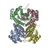
| ||||||||||||||||
|---|---|---|---|---|---|---|---|---|---|---|---|---|---|---|---|---|---|
| 1 |
| ||||||||||||||||
| Unit cell |
| ||||||||||||||||
| Noncrystallographic symmetry (NCS) | NCS oper:
|
-Components
| #1: Protein | Mass: 28167.961 Da / Num. of mol.: 4 Source method: isolated from a genetically manipulated source Source: (gene. exp.) PSEUDOMONAS AERUGINOSA (bacteria) / Strain: PAO1 / Plasmid: PNIC-PA2967 / Production host: References: UniProt: O54438, 3-oxoacyl-[acyl-carrier-protein] reductase #2: Chemical |   Mass: 312.409 Da / Num. of mol.: 2 / Source method: obtained synthetically / Formula: C16H12N2OS2 Mass: 312.409 Da / Num. of mol.: 2 / Source method: obtained synthetically / Formula: C16H12N2OS2#3: Water | ChemComp-HOH / |  Mass: 18.015 Da / Num. of mol.: 25 / Source method: isolated from a natural source / Formula: H2O Mass: 18.015 Da / Num. of mol.: 25 / Source method: isolated from a natural source / Formula: H2ONonpolymer details | 36P: THE LIGAND 2,3-DIHYDROINDOL-1-YL-(2-THIOPHEN-3-YL-1,3- THIAZOL-4-YL)METHANONE IS BOUND AT THE ...36P: THE LIGAND 2,3-DIHYDROIND | |
|---|
-Experimental details
-Experiment
| Experiment | Method: X-RAY DIFFRACTION / Number of used crystals: 1 |
|---|
- Sample preparation
| Crystal | Density Matthews: 1.95 Å3/Da / Density % sol: 36.81 % / Description: NONE |
|---|---|
| Crystal grow | pH: 7 Details: 0.24 M SODIUM MALONATE PH 7.0, 20% (W/V) PEG3350, 1 MM 2,3-DIHYDROINDOL-1-YL-(2-THIOPHEN- 3-YL-1,3-THIAZOL-4-YL)METHANONE, FINAL PROTEIN CONCENTRATION 5 MG/ML |
-Data collection
| Diffraction | Mean temperature: 100 K |
|---|---|
| Diffraction source | Source: SYNCHROTRON / Site: MAX II  / Beamline: I911-2 / Wavelength: 1.0409 / Beamline: I911-2 / Wavelength: 1.0409 |
| Detector | Type: MARRESEARCH / Detector: CCD / Date: Sep 6, 2012 / Details: MIRRORS |
| Radiation | Monochromator: SI(111) / Protocol: SINGLE WAVELENGTH / Monochromatic (M) / Laue (L): M / Scattering type: x-ray |
| Radiation wavelength | Wavelength: 1.0409 Å / Relative weight: 1 |
| Reflection | Resolution: 2.8→15.93 Å / Num. obs: 19603 / % possible obs: 88.2 % / Observed criterion σ(I): -3 / Redundancy: 4.4 % / Biso Wilson estimate: 34.7 Å2 / Rmerge(I) obs: 0.1 / Net I/σ(I): 10.5 |
| Reflection shell | Resolution: 2.8→2.97 Å / Redundancy: 4.6 % / Rmerge(I) obs: 0.4 / Mean I/σ(I) obs: 3.7 / % possible all: 79.9 |
- Processing
| Software |
| ||||||||||||||||||||||||||||||||||||||||||||||||||||||||||||||||||||||||||||||||||||||||||||||||||||||||||||||||||||||||||||||||||||||||||||||||||||||||||||||||||||||||||||||||||||||
|---|---|---|---|---|---|---|---|---|---|---|---|---|---|---|---|---|---|---|---|---|---|---|---|---|---|---|---|---|---|---|---|---|---|---|---|---|---|---|---|---|---|---|---|---|---|---|---|---|---|---|---|---|---|---|---|---|---|---|---|---|---|---|---|---|---|---|---|---|---|---|---|---|---|---|---|---|---|---|---|---|---|---|---|---|---|---|---|---|---|---|---|---|---|---|---|---|---|---|---|---|---|---|---|---|---|---|---|---|---|---|---|---|---|---|---|---|---|---|---|---|---|---|---|---|---|---|---|---|---|---|---|---|---|---|---|---|---|---|---|---|---|---|---|---|---|---|---|---|---|---|---|---|---|---|---|---|---|---|---|---|---|---|---|---|---|---|---|---|---|---|---|---|---|---|---|---|---|---|---|---|---|---|---|
| Refinement | Method to determine structure: MOLECULAR REPLACEMENT Starting model: PDB ENTRY 4BNW, LIGAND-FREE Resolution: 2.8→15.94 Å / Cor.coef. Fo:Fc: 0.925 / Cor.coef. Fo:Fc free: 0.901 / SU B: 33.919 / SU ML: 0.313 / Cross valid method: THROUGHOUT / ESU R Free: 0.468 / Stereochemistry target values: MAXIMUM LIKELIHOOD / Details: HYDROGENS HAVE BEEN ADDED IN THE RIDING POSITIONS.
| ||||||||||||||||||||||||||||||||||||||||||||||||||||||||||||||||||||||||||||||||||||||||||||||||||||||||||||||||||||||||||||||||||||||||||||||||||||||||||||||||||||||||||||||||||||||
| Solvent computation | Ion probe radii: 0.8 Å / Shrinkage radii: 0.8 Å / VDW probe radii: 1.2 Å / Solvent model: BABINET MODEL WITH MASK | ||||||||||||||||||||||||||||||||||||||||||||||||||||||||||||||||||||||||||||||||||||||||||||||||||||||||||||||||||||||||||||||||||||||||||||||||||||||||||||||||||||||||||||||||||||||
| Displacement parameters | Biso mean: 38.305 Å2
| ||||||||||||||||||||||||||||||||||||||||||||||||||||||||||||||||||||||||||||||||||||||||||||||||||||||||||||||||||||||||||||||||||||||||||||||||||||||||||||||||||||||||||||||||||||||
| Refinement step | Cycle: LAST / Resolution: 2.8→15.94 Å
| ||||||||||||||||||||||||||||||||||||||||||||||||||||||||||||||||||||||||||||||||||||||||||||||||||||||||||||||||||||||||||||||||||||||||||||||||||||||||||||||||||||||||||||||||||||||
| Refine LS restraints |
|