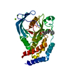
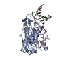
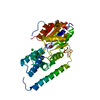
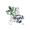
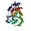
[English] 日本語
Yorodumi
- PDB-4bjo: Nitrate in the active site of PTP1b is a putative mimetic of the ... -

+
Open data


ID or keywords:

Loading...

-
Basic information

Entry
Database: PDB / ID: 4bjo
TitleNitrate in the active site of PTP1b is a putative mimetic of the transition state
ComponentsTYROSINE-PROTEIN PHOSPHATASE NON-RECEPTOR TYPE 1
KeywordsHYDROLASE / PHOSPHATASE / TRANSITION STATE / QUANTUM CHEMISTRY
Function / homology
Function and homology information


regulation of hepatocyte growth factor receptor signaling pathway / PTK6 Down-Regulation / peptidyl-tyrosine dephosphorylation involved in inactivation of protein kinase activity / positive regulation of receptor catabolic process / insulin receptor recycling / negative regulation of PERK-mediated unfolded protein response / negative regulation of vascular endothelial growth factor receptor signaling pathway / regulation of intracellular protein transport / IRE1-mediated unfolded protein response / cytoplasmic side of endoplasmic reticulum membrane ...regulation of hepatocyte growth factor receptor signaling pathway / PTK6 Down-Regulation / peptidyl-tyrosine dephosphorylation involved in inactivation of protein kinase activity / positive regulation of receptor catabolic process / insulin receptor recycling / negative regulation of PERK-mediated unfolded protein response / negative regulation of vascular endothelial growth factor receptor signaling pathway / regulation of intracellular protein transport / IRE1-mediated unfolded protein response / cytoplasmic side of endoplasmic reticulum membrane / sorting endosome / mitochondrial crista / platelet-derived growth factor receptor-beta signaling pathway / positive regulation of IRE1-mediated unfolded protein response / regulation of type I interferon-mediated signaling pathway / regulation of endocytosis / non-membrane spanning protein tyrosine phosphatase activity / peptidyl-tyrosine dephosphorylation / Regulation of IFNA/IFNB signaling / regulation of signal transduction / cellular response to unfolded protein / positive regulation of protein tyrosine kinase activity / growth hormone receptor signaling pathway via JAK-STAT / negative regulation of endoplasmic reticulum stress-induced intrinsic apoptotic signaling pathway / negative regulation of signal transduction / Regulation of IFNG signaling / MECP2 regulates neuronal receptors and channels / endoplasmic reticulum unfolded protein response / Growth hormone receptor signaling / positive regulation of JUN kinase activity / negative regulation of insulin receptor signaling pathway / Insulin receptor recycling / ephrin receptor binding / Integrin signaling / protein dephosphorylation / negative regulation of MAP kinase activity / protein-tyrosine-phosphatase / protein phosphatase 2A binding / protein tyrosine phosphatase activity / endosome lumen / insulin receptor binding / Negative regulation of MET activity / negative regulation of ERK1 and ERK2 cascade / receptor tyrosine kinase binding / insulin receptor signaling pathway / actin cytoskeleton organization / early endosome / mitochondrial matrix / cadherin binding / protein kinase binding / enzyme binding / endoplasmic reticulum / protein-containing complex / RNA binding / zinc ion binding / cytosol / cytoplasm
Similarity search - Function
Protein-tyrosine phosphatase, non-receptor type-1/2 / Protein tyrosine phosphatase superfamily / Protein-Tyrosine Phosphatase; Chain A / Protein tyrosine phosphatase, catalytic domain / PTP type protein phosphatase domain profile. / Protein-tyrosine phosphatase / Tyrosine-specific protein phosphatase, PTPase domain / Protein-tyrosine phosphatase, catalytic / Protein tyrosine phosphatase, catalytic domain motif / Tyrosine specific protein phosphatases active site. ...Protein-tyrosine phosphatase, non-receptor type-1/2 / Protein tyrosine phosphatase superfamily / Protein-Tyrosine Phosphatase; Chain A / Protein tyrosine phosphatase, catalytic domain / PTP type protein phosphatase domain profile. / Protein-tyrosine phosphatase / Tyrosine-specific protein phosphatase, PTPase domain / Protein-tyrosine phosphatase, catalytic / Protein tyrosine phosphatase, catalytic domain motif / Tyrosine specific protein phosphatases active site. / Protein-tyrosine phosphatase, active site / Tyrosine specific protein phosphatases domain profile. / Tyrosine-specific protein phosphatases domain / Protein-tyrosine phosphatase-like / Alpha-Beta Complex / Alpha Beta
Similarity search - Domain/homology
NITRATE ION / Tyrosine-protein phosphatase non-receptor type 1
Similarity search - Component
Biological speciesHOMO SAPIENS (human)
MethodX-RAY DIFFRACTION / SYNCHROTRON / MOLECULAR REPLACEMENT / Resolution: 2.06 Å
AuthorsKenny, P.W. / Newman, J. / Peat, T.S.
CitationJournal: Acta Crystallogr.,Sect.D / Year: 2014
Title: Nitrate in the Active Site of Protein Tyrosine Phosphatase 1B is a Putative Mimetic of the Transition State.
Authors: Kenny, P.W. / Newman, J. / Peat, T.S.
History
DepositionApr 19, 2013Deposition site: PDBE / Processing site: PDBE
Revision 1.0Feb 12, 2014Provider: repository / Type: Initial release
Revision 1.1Feb 26, 2014Group: Database references
Revision 1.2May 8, 2019Group: Data collection / Experimental preparation / Other
Category: exptl_crystal_grow / pdbx_database_proc ...exptl_crystal_grow / pdbx_database_proc / pdbx_database_status / struct_biol
Item: _exptl_crystal_grow.temp / _pdbx_database_status.recvd_author_approval
Revision 1.3Dec 20, 2023Group: Data collection / Database references ...Data collection / Database references / Derived calculations / Other / Refinement description
Category: chem_comp_atom / chem_comp_bond ...chem_comp_atom / chem_comp_bond / database_2 / pdbx_database_status / pdbx_initial_refinement_model / struct_ncs_dom_lim / struct_site
Item: _database_2.pdbx_DOI / _database_2.pdbx_database_accession ..._database_2.pdbx_DOI / _database_2.pdbx_database_accession / _pdbx_database_status.status_code_sf / _struct_ncs_dom_lim.beg_auth_comp_id / _struct_ncs_dom_lim.beg_label_asym_id / _struct_ncs_dom_lim.beg_label_comp_id / _struct_ncs_dom_lim.beg_label_seq_id / _struct_ncs_dom_lim.end_auth_comp_id / _struct_ncs_dom_lim.end_label_asym_id / _struct_ncs_dom_lim.end_label_comp_id / _struct_ncs_dom_lim.end_label_seq_id / _struct_site.pdbx_auth_asym_id / _struct_site.pdbx_auth_comp_id / _struct_site.pdbx_auth_seq_id

-
Structure visualization

Structure viewerMolecule:
MolmilJmol/JSmol

Downloads & links

-
Assembly

Deposited unit
A: TYROSINE-PROTEIN PHOSPHATASE NON-RECEPTOR TYPE 1
B: TYROSINE-PROTEIN PHOSPHATASE NON-RECEPTOR TYPE 1
hetero molecules


Theoretical massNumber of molelcules
Total (without water)79,07411
Polymers78,6972
Non-polymers3779
Water2,234124
1
A: TYROSINE-PROTEIN PHOSPHATASE NON-RECEPTOR TYPE 1
hetero molecules


Theoretical massNumber of molelcules
Total (without water)39,5927
Polymers39,3491
Non-polymers2446
Water181
TypeNameSymmetry operationNumber
identity operation1_555x,y,z1
MethodPISA
2
B: TYROSINE-PROTEIN PHOSPHATASE NON-RECEPTOR TYPE 1
hetero molecules


Theoretical massNumber of molelcules
Total (without water)39,4824
Polymers39,3491
Non-polymers1333
Water181
TypeNameSymmetry operationNumber
identity operation1_555x,y,z1
MethodPISA
Unit cell
Length a, b, c (Å)161.620, 66.765, 68.771
Angle α, β, γ (deg.)90.00, 95.01, 90.00
Int Tables number5
Space group name H-MC121
Noncrystallographic symmetry (NCS)NCS domain:
IDEns-IDDetails
11A
21B

NCS domain segments:

Component-ID: 0 / Ens-ID: 1 / Beg auth comp-ID: ASN / Beg label comp-ID: ASN / End auth comp-ID: PHE / End label comp-ID: PHE / Refine code: 0 / Auth seq-ID: -5 - 280 / Label seq-ID: 12 - 297

Dom-IDAuth asym-IDLabel asym-ID
1AA
2BB

-
Components

-
Protein , 1 types, 2 molecules AB

#1: Protein TYROSINE-PROTEIN PHOSPHATASE NON-RECEPTOR TYPE 1 / PTP1B PROTEIN PHOSPHATASE / PROTEIN-TYROSINE PHOSPHATASE 1B


Mass: 39348.691 Da / Num. of mol.: 2 / Fragment: CATALYTIC DOMAIN, RESIDUES 2-321
Source method: isolated from a genetically manipulated source
Source: (gene. exp.) HOMO SAPIENS (human) / Production host: ESCHERICHIA COLI (E. coli) / Strain (production host): BL21(DE3) / References: UniProt: P18031, protein-tyrosine-phosphatase

-
Non-polymers , 5 types, 133 molecules

#2: Chemical
ChemComp-CL / CHLORIDE ION / Chloride


Mass: 35.453 Da / Num. of mol.: 4 / Source method: obtained synthetically / Formula: Cl
#3: Chemical ChemComp-MG / MAGNESIUM ION


Mass: 24.305 Da / Num. of mol.: 2 / Source method: obtained synthetically / Formula: Mg
#4: Chemical ChemComp-NO3 / NITRATE ION / Nitrate


Mass: 62.005 Da / Num. of mol.: 2 / Source method: obtained synthetically / Formula: NO3
#5: Chemical ChemComp-EDO / 1,2-ETHANEDIOL / ETHYLENE GLYCOL / Ethylene glycol


Mass: 62.068 Da / Num. of mol.: 1 / Source method: obtained synthetically / Formula: C2H6O2
#6: Water ChemComp-HOH / water / Water


Mass: 18.015 Da / Num. of mol.: 124 / Source method: isolated from a natural source / Formula: H2O

-
Details

Sequence detailsHIS-TAG AT N-TERMINUS

-
Experimental details

-
Experiment

ExperimentMethod: X-RAY DIFFRACTION / Number of used crystals: 1

-
Sample preparation

CrystalDensity Matthews: 2.79 Å3/Da / Density % sol: 55.9 % / Description: NONE
Crystal growTemperature: 281 K / pH: 8.1
Details: 100 MM HEPES PH 8.1, 25.4% PEG 4000, 225 MM MGNO3 AT 281K.

-
Data collection

DiffractionMean temperature: 100 K
Diffraction sourceSource: SYNCHROTRON / Site: Australian Synchrotron / Beamline: MX1 / Wavelength: 1.033
DetectorType: ADSC CCD / Detector: CCD / Date: Feb 16, 2012 / Details: MIRRORS
RadiationProtocol: SINGLE WAVELENGTH / Monochromatic (M) / Laue (L): M / Scattering type: x-ray
Radiation wavelengthWavelength: 1.033 Å / Relative weight: 1
ReflectionResolution: 2.06→80.5 Å / Num. obs: 44854 / % possible obs: 99 % / Observed criterion σ(I): 0 / Redundancy: 7.1 % / Rmerge(I) obs: 0.05 / Net I/σ(I): 23.6
Reflection shellResolution: 2.06→2.17 Å / Redundancy: 4.6 % / Rmerge(I) obs: 0.61 / Mean I/σ(I) obs: 2.6 / % possible all: 93.5

-
Processing

Software
NameVersionClassification
REFMAC5.7.0032refinement
XDSdata reduction
SCALAdata scaling
PHASERphasing
RefinementMethod to determine structure: MOLECULAR REPLACEMENT
Starting model: PDB ENTRY 3AJ5
Resolution: 2.06→68.6 Å / Cor.coef. Fo:Fc: 0.955 / Cor.coef. Fo:Fc free: 0.947 / SU B: 5.475 / SU ML: 0.142 / Cross valid method: THROUGHOUT / ESU R: 0.208 / ESU R Free: 0.17 / Stereochemistry target values: MAXIMUM LIKELIHOOD
Details: HYDROGENS HAVE BEEN ADDED IN THE RIDING POSITIONS. U VALUES REFINED INDIVIDUALLY
RfactorNum. reflection% reflectionSelection details
Rfree0.23932 2290 5.1 %RANDOM
Rwork0.21529 ---
obs0.21652 42525 98.88 %-
Solvent computationIon probe radii: 0.8 Å / Shrinkage radii: 0.8 Å / VDW probe radii: 1.2 Å / Solvent model: MASK
Displacement parametersBiso mean: 49.386 Å2
Baniso -1Baniso -2Baniso -3
1--2.19 Å20 Å20.07 Å2
2--2.15 Å20 Å2
3---0.03 Å2
Refinement stepCycle: LAST / Resolution: 2.06→68.6 Å
ProteinNucleic acidLigandSolventTotal
Num. atoms4671 0 18 124 4813
Refine LS restraints
Refine-IDTypeDev idealDev ideal targetNumber
X-RAY DIFFRACTIONr_bond_refined_d0.0090.0194846
X-RAY DIFFRACTIONr_bond_other_d0.0030.024582
X-RAY DIFFRACTIONr_angle_refined_deg1.2351.9566527
X-RAY DIFFRACTIONr_angle_other_deg0.901310578
X-RAY DIFFRACTIONr_dihedral_angle_1_deg5.6375578
X-RAY DIFFRACTIONr_dihedral_angle_2_deg32.5323.875240
X-RAY DIFFRACTIONr_dihedral_angle_3_deg14.56715897
X-RAY DIFFRACTIONr_dihedral_angle_4_deg17.4251535
X-RAY DIFFRACTIONr_chiral_restr0.0680.2690
X-RAY DIFFRACTIONr_gen_planes_refined0.0060.0215433
X-RAY DIFFRACTIONr_gen_planes_other0.0030.021146
X-RAY DIFFRACTIONr_nbd_refined
X-RAY DIFFRACTIONr_nbd_other
X-RAY DIFFRACTIONr_nbtor_refined
X-RAY DIFFRACTIONr_nbtor_other
X-RAY DIFFRACTIONr_xyhbond_nbd_refined
X-RAY DIFFRACTIONr_xyhbond_nbd_other
X-RAY DIFFRACTIONr_metal_ion_refined
X-RAY DIFFRACTIONr_metal_ion_other
X-RAY DIFFRACTIONr_symmetry_vdw_refined
X-RAY DIFFRACTIONr_symmetry_vdw_other
X-RAY DIFFRACTIONr_symmetry_hbond_refined
X-RAY DIFFRACTIONr_symmetry_hbond_other
X-RAY DIFFRACTIONr_symmetry_metal_ion_refined
X-RAY DIFFRACTIONr_symmetry_metal_ion_other
X-RAY DIFFRACTIONr_mcbond_it3.8536.3042306
X-RAY DIFFRACTIONr_mcbond_other3.8516.3022305
X-RAY DIFFRACTIONr_mcangle_it5.39410.6052886
X-RAY DIFFRACTIONr_mcangle_other5.39410.6082887
X-RAY DIFFRACTIONr_scbond_it4.0786.8392539
X-RAY DIFFRACTIONr_scbond_other4.0766.8392539
X-RAY DIFFRACTIONr_scangle_it
X-RAY DIFFRACTIONr_scangle_other6.25711.2953642
X-RAY DIFFRACTIONr_long_range_B_refined8.15633.685569
X-RAY DIFFRACTIONr_long_range_B_other8.15533.6815570
X-RAY DIFFRACTIONr_rigid_bond_restr
X-RAY DIFFRACTIONr_sphericity_free
X-RAY DIFFRACTIONr_sphericity_bonded
Refine LS restraints NCS

Ens-ID: 1 / Number: 17493 / Refine-ID: X-RAY DIFFRACTION / Type: interatomic distance / Rms dev position: 0.12 Å / Weight position: 0.05

Dom-IDAuth asym-ID
1A
2B
LS refinement shellResolution: 2.06→2.113 Å / Total num. of bins used: 20
RfactorNum. reflection% reflection
Rfree0.402 179 -
Rwork0.334 2764 -
obs--89.32 %

+
About Yorodumi

-
News

-
Feb 9, 2022. New format data for meta-information of EMDB entries

New format data for meta-information of EMDB entries

  • Version 3 of the EMDB header file is now the official format.
  • The previous official version 1.9 will be removed from the archive.

Related info.:EMDB header

External links:wwPDB to switch to version 3 of the EMDB data model

-
Aug 12, 2020. Covid-19 info

Covid-19 info

URL: https://pdbj.org/emnavi/covid19.php

New page: Covid-19 featured information page in EM Navigator.

Related info.:Covid-19 info / Mar 5, 2020. Novel coronavirus structure data

+
Mar 5, 2020. Novel coronavirus structure data

Novel coronavirus structure data

Related info.:Yorodumi Speices / Aug 12, 2020. Covid-19 info

External links:COVID-19 featured content - PDBj / Molecule of the Month (242):Coronavirus Proteases

+
Jan 31, 2019. EMDB accession codes are about to change! (news from PDBe EMDB page)

EMDB accession codes are about to change! (news from PDBe EMDB page)

  • The allocation of 4 digits for EMDB accession codes will soon come to an end. Whilst these codes will remain in use, new EMDB accession codes will include an additional digit and will expand incrementally as the available range of codes is exhausted. The current 4-digit format prefixed with “EMD-” (i.e. EMD-XXXX) will advance to a 5-digit format (i.e. EMD-XXXXX), and so on. It is currently estimated that the 4-digit codes will be depleted around Spring 2019, at which point the 5-digit format will come into force.
  • The EM Navigator/Yorodumi systems omit the EMD- prefix.

Related info.:Q: What is EMD? / ID/Accession-code notation in Yorodumi/EM Navigator

External links:EMDB Accession Codes are Changing Soon! / Contact to PDBj

+
Jul 12, 2017. Major update of PDB

Major update of PDB

  • wwPDB released updated PDB data conforming to the new PDBx/mmCIF dictionary.
  • This is a major update changing the version number from 4 to 5, and with Remediation, in which all the entries are updated.
  • In this update, many items about electron microscopy experimental information are reorganized (e.g. em_software).
  • Now, EM Navigator and Yorodumi are based on the updated data.

External links:wwPDB Remediation / Enriched Model Files Conforming to OneDep Data Standards Now Available in the PDB FTP Archive

-
Yorodumi

Thousand views of thousand structures

  • Yorodumi is a browser for structure data from EMDB, PDB, SASBDB, etc.
  • This page is also the successor to EM Navigator detail page, and also detail information page/front-end page for Omokage search.
  • The word "yorodu" (or yorozu) is an old Japanese word meaning "ten thousand". "mi" (miru) is to see.

Related info.:EMDB / PDB / SASBDB / Comparison of 3 databanks / Yorodumi Search / Aug 31, 2016. New EM Navigator & Yorodumi / Yorodumi Papers / Jmol/JSmol / Function and homology information / Changes in new EM Navigator and Yorodumi

Read more