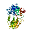
- PDB-4b4a: Structure of the TatC core of the twin arginine protein transloca... -
+
Open data
ID or keywords:
Loading...
-
Basic information
Entry
Database: PDB / ID: 4b4a
Title
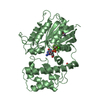
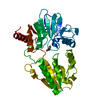
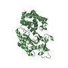
Structure of the TatC core of the twin arginine protein translocation system
Components
SEC-INDEPENDENT PROTEIN TRANSLOCASE PROTEIN TATC
Keywords
TRANSPORT PROTEIN / TAT SECRETION SYSTEM / PROTEIN TRANSLOCATION
Function / homology
Sec-independent periplasmic protein translocase, conserved site / TatC family signature. / Sec-independent periplasmic protein translocase TatC / Sec-independent protein translocase protein (TatC) / proton motive force dependent protein transmembrane transporter activity / TAT protein transport complex / protein transport by the Tat complex / intracellular protein transmembrane transport / Sec-independent protein translocase protein TatC
Function and homology information
Biological species
AQUIFEX AEOLICUS (bacteria)
Method
X-RAY DIFFRACTION / SYNCHROTRON / SAD / Resolution: 3.5 Å
Protocol: SINGLE WAVELENGTH / Monochromatic (M) / Laue (L): M / Scattering type: x-ray
Radiation wavelength
Wavelength: 0.92 Å / Relative weight: 1
Reflection
Resolution: 3.5→15 Å / Num. obs: 7820 / % possible obs: 96.8 % / Observed criterion σ(I): 0 / Redundancy: 3.3 % / Biso Wilson estimate: 132.26 Å2 / Rmerge(I) obs: 0.03 / Net I/σ(I): 13.9
Reflection shell
Resolution: 3.5→3.6 Å / Redundancy: 3.4 % / Rmerge(I) obs: 0.95 / Mean I/σ(I) obs: 1.4 / % possible all: 96.6
-
Processing
Software
Name
Version
Classification
XDS
THROUGHXIA2
datareduction
Aimless
THROUGHXIA2
datascaling
PHENIX.HYSS
phasing
autoSHARP
phasing
BUSTER
2.11.2
refinement
Refinement
Method to determine structure: SAD Starting model: NONE Resolution: 3.5→22 Å / Cor.coef. Fo:Fc: 0.8869 / Cor.coef. Fo:Fc free: 0.8553 / SU R Cruickshank DPI: 4.314 / Cross valid method: THROUGHOUT / σ(F): 0 / SU R Blow DPI: 1.44 / SU Rfree Blow DPI: 0.479 / SU Rfree Cruickshank DPI: 0.497 Details: IDEAL-DIST CONTACT TERM CONTACT SETUP. ALL ATOMS HAVE CCP4 ATOM TYPE FROM LIBRARY
Rfactor
Num. reflection
% reflection
Selection details
Rfree
0.2882
386
4.89 %
RANDOM
Rwork
0.2518
-
-
-
obs
0.2535
7900
96.29 %
-
Displacement parameters
Biso mean: 159.38 Å2
Baniso -1
Baniso -2
Baniso -3
1-
-18.3276 Å2
0 Å2
0 Å2
2-
-
-18.3276 Å2
0 Å2
3-
-
-
36.6553 Å2
Refine analyze
Luzzati coordinate error obs: 0.92 Å
Refinement step
Cycle: LAST / Resolution: 3.5→22 Å
Protein
Nucleic acid
Ligand
Solvent
Total
Num. atoms
1804
0
69
0
1873
Refine LS restraints
Refine-ID
Type
Dev ideal
Number
Restraint function
Weight
X-RAY DIFFRACTION
t_bond_d
0.01
2007
HARMONIC
2
X-RAY DIFFRACTION
t_angle_deg
1.27
2808
HARMONIC
2
X-RAY DIFFRACTION
t_dihedral_angle_d
670
SINUSOIDAL
2
X-RAY DIFFRACTION
t_incorr_chiral_ct
X-RAY DIFFRACTION
t_pseud_angle
X-RAY DIFFRACTION
t_trig_c_planes
19
HARMONIC
2
X-RAY DIFFRACTION
t_gen_planes
266
HARMONIC
5
X-RAY DIFFRACTION
t_it
2007
HARMONIC
20
X-RAY DIFFRACTION
t_nbd
X-RAY DIFFRACTION
t_omega_torsion
2.6
X-RAY DIFFRACTION
t_other_torsion
24.18
X-RAY DIFFRACTION
t_improper_torsion
X-RAY DIFFRACTION
t_chiral_improper_torsion
267
SEMIHARMONIC
5
X-RAY DIFFRACTION
t_sum_occupancies
X-RAY DIFFRACTION
t_utility_distance
X-RAY DIFFRACTION
t_utility_angle
X-RAY DIFFRACTION
t_utility_torsion
X-RAY DIFFRACTION
t_ideal_dist_contact
2308
SEMIHARMONIC
4
LS refinement shell
Resolution: 3.5→3.91 Å / Total num. of bins used: 5
Rfactor
Num. reflection
% reflection
Rfree
0.2484
126
5.78 %
Rwork
0.2399
2053
-
all
0.2404
2179
-
obs
-
-
96.29 %
+
About Yorodumi
-
News
-
Feb 9, 2022. New format data for meta-information of EMDB entries
New format data for meta-information of EMDB entries
Version 3 of the EMDB header file is now the official format.
The previous official version 1.9 will be removed from the archive.
In the structure databanks used in Yorodumi, some data are registered as the other names, "COVID-19 virus" and "2019-nCoV". Here are the details of the virus and the list of structure data.
Jan 31, 2019. EMDB accession codes are about to change! (news from PDBe EMDB page)
EMDB accession codes are about to change! (news from PDBe EMDB page)
The allocation of 4 digits for EMDB accession codes will soon come to an end. Whilst these codes will remain in use, new EMDB accession codes will include an additional digit and will expand incrementally as the available range of codes is exhausted. The current 4-digit format prefixed with “EMD-” (i.e. EMD-XXXX) will advance to a 5-digit format (i.e. EMD-XXXXX), and so on. It is currently estimated that the 4-digit codes will be depleted around Spring 2019, at which point the 5-digit format will come into force.
The EM Navigator/Yorodumi systems omit the EMD- prefix.
Related info.:Q: What is EMD? / ID/Accession-code notation in Yorodumi/EM Navigator
Yorodumi is a browser for structure data from EMDB, PDB, SASBDB, etc.
This page is also the successor to EM Navigator detail page, and also detail information page/front-end page for Omokage search.
The word "yorodu" (or yorozu) is an old Japanese word meaning "ten thousand". "mi" (miru) is to see.
Related info.:EMDB / PDB / SASBDB / Comparison of 3 databanks / Yorodumi Search / Aug 31, 2016. New EM Navigator & Yorodumi / Yorodumi Papers / Jmol/JSmol / Function and homology information / Changes in new EM Navigator and Yorodumi
 Movie
Movie Controller

 Yorodumi
 Open data
 Basic information
 Components
 Keywords
 Function and homology information
 AQUIFEX AEOLICUS (bacteria)
 X-RAY DIFFRACTION /
  Authors
 Citation
 Structure visualization
 Downloads & links
 Other downloads
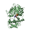


 PDBj
PDBj Assembly
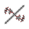
 Mass: 1005.188 Da / Num. of mol.: 1 / Source method: obtained synthetically / Formula: C47H88O22 / Comment: detergent*YM
 Sample preparation
 / Beamline: I04-1 / Wavelength: 0.92
  Processing