 Movie
Movie Controller

[English] 日本語
 Yorodumi
- PDB-4ax2: New Type VI-secreted toxins and self-resistance proteins in Serra... -
+ Open data

- Basic information
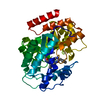
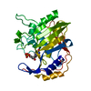
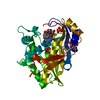
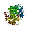
| Entry | Database: PDB / ID: 4ax2 | ||||||
|---|---|---|---|---|---|---|---|
| Title | New Type VI-secreted toxins and self-resistance proteins in Serratia marcescens | ||||||
 Components Components | RAP1B | ||||||
 Keywords Keywords | TOXIN / RESISTANCE PROTEIN / HELICAL FOLD / S-SAD PHASING | ||||||
| Function / homology | Type VI secretion system (T6SS), amidase immunity protein / T6SS superfamily / Type VI secretion system (T6SS), amidase immunity protein / Four Helix Bundle (Hemerythrin (Met), subunit A) - #1620 / Four Helix Bundle (Hemerythrin (Met), subunit A) / Up-down Bundle / Mainly Alpha / IODIDE ION / Rap1b Function and homology information Function and homology information | ||||||
| Biological species |  SERRATIA MARCESCENS (bacteria) SERRATIA MARCESCENS (bacteria) | ||||||
| Method |  X-RAY DIFFRACTION / SAD / Resolution: 1.88 Å X-RAY DIFFRACTION / SAD / Resolution: 1.88 Å | ||||||
 Authors Authors | English, G. / Trunk, K. / Rao, V.A. / Srikannathasan, V. / Fritsch, M.J. / Guo, M. / Hunter, W.N. / Coulthurst, S.J. | ||||||
 Citation Citation | Journal: Mol.Microbiol. / Year: 2012 Title: New Secreted Toxins and Immunity Proteins Encoded within the Type Vi Secretion System Gene Cluster of Serratia Marcescens Authors: English, G. / Trunk, K. / Rao, V.A. / Srikannathasan, V. / Hunter, W.N. / Coulthurst, S.J. | ||||||
| History |
|
- Structure visualization
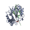
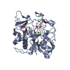
| Structure viewer | Molecule: MolmilJmol/JSmol |
|---|
- Downloads & links
-Download
| PDBx/mmCIF format | 4ax2.cif.gz | 69 KB | Display | PDBx/mmCIF format |
|---|---|---|---|---|
| PDB format | pdb4ax2.ent.gz | 51.5 KB | Display | PDB format |
| PDBx/mmJSON format | 4ax2.json.gz | Tree view | PDBx/mmJSON format | |
| Others |  Other downloads Other downloads |
-Validation report
| Arichive directory | https://data.pdbj.org/pub/pdb/validation_reports/ax/4ax2ftp://data.pdbj.org/pub/pdb/validation_reports/ax/4ax2 | HTTPS FTP |
|---|
-Related structure data


-Links
 PDBj
PDBj- Assembly
| Deposited unit | 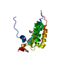
| ||||||||
|---|---|---|---|---|---|---|---|---|---|
| 1 | 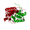
| ||||||||
| Unit cell |
|
-Components
| #1: Protein | Mass: 16364.356 Da / Num. of mol.: 1 Source method: isolated from a genetically manipulated source Source: (gene. exp.) SERRATIA MARCESCENS (bacteria) / Strain: DB10 / Plasmid: PET15BTEV_PSC503 / Production host: | ||||||||||
|---|---|---|---|---|---|---|---|---|---|---|---|
| #2: Chemical |   Mass: 126.904 Da / Num. of mol.: 2 / Source method: obtained synthetically / Formula: I Mass: 126.904 Da / Num. of mol.: 2 / Source method: obtained synthetically / Formula: I#3: Chemical | ChemComp-EDO /   Mass: 62.068 Da / Num. of mol.: 5 / Source method: obtained synthetically / Formula: C2H6O2 Mass: 62.068 Da / Num. of mol.: 5 / Source method: obtained synthetically / Formula: C2H6O2#4: Water | ChemComp-HOH / |  Mass: 18.015 Da / Num. of mol.: 111 / Source method: isolated from a natural source / Formula: H2O Mass: 18.015 Da / Num. of mol.: 111 / Source method: isolated from a natural source / Formula: H2OHas protein modification | Y | Nonpolymer details | IODIDE ION (IOD): HALIDE SOAK. IODIDES USED FOR SAD PHASING ALONG WITH S ATOMS. | Sequence details | GENOME IS NOT YET IN ANY DATABASE | |
-Experimental details
-Experiment
| Experiment | Method: X-RAY DIFFRACTION / Number of used crystals: 1 |
|---|
- Sample preparation
| Crystal | Density Matthews: 2.51 Å3/Da / Density % sol: 51 % / Description: NONE |
|---|---|
| Crystal grow | pH: 7.5 / Details: 0.55M LI2SO4, 5% PEG 8000, pH 7.5 |
-Data collection
| Diffraction | Mean temperature: 93 K |
|---|---|
| Diffraction source | Source: ROTATING ANODE / Type: RIGAKU MICROMAX-007 / Wavelength: 1.5418 |
| Detector | Type: RIGAKU IMAGE PLATE / Detector: IMAGE PLATE / Date: Mar 24, 2011 / Details: MIRRORS |
| Radiation | Monochromator: SILICON / Protocol: SINGLE WAVELENGTH / Monochromatic (M) / Laue (L): M / Scattering type: x-ray |
| Radiation wavelength | Wavelength: 1.5418 Å / Relative weight: 1 |
| Reflection | Resolution: 1.88→67.5 Å / Num. obs: 14813 / % possible obs: 100 % / Observed criterion σ(I): 0 / Redundancy: 11 % / Biso Wilson estimate: 28.3 Å2 / Rmerge(I) obs: 0.05 / Net I/σ(I): 26.6 |
| Reflection shell | Resolution: 1.88→1.98 Å / Redundancy: 10.2 % / Rmerge(I) obs: 0.38 / Mean I/σ(I) obs: 5.9 / % possible all: 100 |
- Processing
| Software |
| ||||||||||||||||||||||||||||||||||||||||||||||||||||||||||||||||||||||||||||||||||||||||||||||||||||||||||||||||||||||||||||||||||||||||||||||||||||||||||||||||||||||||||||||||||||||
|---|---|---|---|---|---|---|---|---|---|---|---|---|---|---|---|---|---|---|---|---|---|---|---|---|---|---|---|---|---|---|---|---|---|---|---|---|---|---|---|---|---|---|---|---|---|---|---|---|---|---|---|---|---|---|---|---|---|---|---|---|---|---|---|---|---|---|---|---|---|---|---|---|---|---|---|---|---|---|---|---|---|---|---|---|---|---|---|---|---|---|---|---|---|---|---|---|---|---|---|---|---|---|---|---|---|---|---|---|---|---|---|---|---|---|---|---|---|---|---|---|---|---|---|---|---|---|---|---|---|---|---|---|---|---|---|---|---|---|---|---|---|---|---|---|---|---|---|---|---|---|---|---|---|---|---|---|---|---|---|---|---|---|---|---|---|---|---|---|---|---|---|---|---|---|---|---|---|---|---|---|---|---|---|
| Refinement | Method to determine structure: SAD Starting model: NONE Resolution: 1.88→67.52 Å / Cor.coef. Fo:Fc: 0.956 / Cor.coef. Fo:Fc free: 0.929 / SU B: 4.609 / SU ML: 0.072 / Cross valid method: THROUGHOUT / ESU R: 0.123 / ESU R Free: 0.122 / Stereochemistry target values: MAXIMUM LIKELIHOOD / Details: HYDROGENS HAVE BEEN ADDED IN THE RIDING POSITIONS.
| ||||||||||||||||||||||||||||||||||||||||||||||||||||||||||||||||||||||||||||||||||||||||||||||||||||||||||||||||||||||||||||||||||||||||||||||||||||||||||||||||||||||||||||||||||||||
| Solvent computation | Ion probe radii: 0.8 Å / Shrinkage radii: 0.8 Å / VDW probe radii: 1.2 Å / Solvent model: MASK | ||||||||||||||||||||||||||||||||||||||||||||||||||||||||||||||||||||||||||||||||||||||||||||||||||||||||||||||||||||||||||||||||||||||||||||||||||||||||||||||||||||||||||||||||||||||
| Displacement parameters | Biso mean: 21.1 Å2
| ||||||||||||||||||||||||||||||||||||||||||||||||||||||||||||||||||||||||||||||||||||||||||||||||||||||||||||||||||||||||||||||||||||||||||||||||||||||||||||||||||||||||||||||||||||||
| Refinement step | Cycle: LAST / Resolution: 1.88→67.52 Å
| ||||||||||||||||||||||||||||||||||||||||||||||||||||||||||||||||||||||||||||||||||||||||||||||||||||||||||||||||||||||||||||||||||||||||||||||||||||||||||||||||||||||||||||||||||||||
| Refine LS restraints |
|