 Movie
Movie Controller

[English] 日本語
 Yorodumi
- PDB-4at9: Crystal structure of the NF90-NF45 dimerisation domain complex wi... -
+ Open data

- Basic information
| Entry | Database: PDB / ID: 4at9 | ||||||
|---|---|---|---|---|---|---|---|
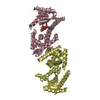
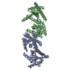
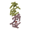
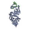
| Title | Crystal structure of the NF90-NF45 dimerisation domain complex with UTP | ||||||
 Components Components |
| ||||||
 Keywords Keywords | IMMUNE SYSTEM / DRPB76 / NFAR / ILF3 / ILF2 / TEMPLATE-FREE NUCLEOTIDYLTRANSFERASE FOLD | ||||||
| Function / homology |  Function and homology information Function and homology informationRegulation of CDH11 gene transcription / spliceosome-depend formation of circular RNA / PKR-mediated signaling / mRNA 3'-UTR AU-rich region binding / negative regulation of viral genome replication / Neutrophil degranulation / double-stranded RNA binding / virus receptor activity / defense response to virus / protein phosphorylation ...Regulation of CDH11 gene transcription / spliceosome-depend formation of circular RNA / PKR-mediated signaling / mRNA 3'-UTR AU-rich region binding / negative regulation of viral genome replication / Neutrophil degranulation / double-stranded RNA binding / virus receptor activity / defense response to virus / protein phosphorylation / negative regulation of translation / single-stranded RNA binding / ribonucleoprotein complex / negative regulation of DNA-templated transcription / positive regulation of DNA-templated transcription / nucleolus / enzyme binding / mitochondrion / DNA binding / nucleoplasm / nucleus / cytoplasm / cytosol Similarity search - Function | ||||||
| Biological species |  | ||||||
| Method |  X-RAY DIFFRACTION / SYNCHROTRON / MOLECULAR REPLACEMENT / Resolution: 2.803 Å X-RAY DIFFRACTION / SYNCHROTRON / MOLECULAR REPLACEMENT / Resolution: 2.803 Å | ||||||
 Authors Authors | Wolkowicz, U.M. / Cook, A.G. | ||||||
 Citation Citation | Journal: Nucleic Acids Res. / Year: 2012 Title: NF45 Dimerizes with NF90, Zfr and Spnr Via a Conserved Domain that Has a Nucleotidyltransferase Fold. Authors: Wolkowicz, U.M. / Cook, A.G. | ||||||
| History |
|
- Structure visualization
| Structure viewer | Molecule: MolmilJmol/JSmol |
|---|
- Downloads & links
-Download
| PDBx/mmCIF format | 4at9.cif.gz | 126.2 KB | Display | PDBx/mmCIF format |
|---|---|---|---|---|
| PDB format | pdb4at9.ent.gz | 95.5 KB | Display | PDB format |
| PDBx/mmJSON format | 4at9.json.gz | Tree view | PDBx/mmJSON format | |
| Others |  Other downloads Other downloads |
-Validation report
| Arichive directory | https://data.pdbj.org/pub/pdb/validation_reports/at/4at9ftp://data.pdbj.org/pub/pdb/validation_reports/at/4at9 | HTTPS FTP |
|---|
-Related structure data
| Related structure data | 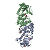 4at7SC 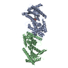 4at8C  4atbC S: Starting model for refinement C: citing same article ( |
|---|---|
| Similar structure data |


-Links
 PDBj
PDBj


- Assembly
| Deposited unit | 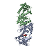
| ||||||||
|---|---|---|---|---|---|---|---|---|---|
| 1 |
| ||||||||
| Unit cell |
|
-Components
| #1: Protein | Mass: 40359.840 Da / Num. of mol.: 1 / Fragment: DZF DOMAIN, RESIDUES 29-390 Source method: isolated from a genetically manipulated source Source: (gene. exp.)  |
|---|---|
| #2: Protein | Mass: 42526.727 Da / Num. of mol.: 1 / Fragment: DZF DOMAIN, RESIDUES 1-381 Source method: isolated from a genetically manipulated source Source: (gene. exp.) |
| #3: Chemical | ChemComp-UTP /   Mass: 484.141 Da / Num. of mol.: 1 / Source method: obtained synthetically / Formula: C9H15N2O15P3 / Comment: UTP*YM Mass: 484.141 Da / Num. of mol.: 1 / Source method: obtained synthetically / Formula: C9H15N2O15P3 / Comment: UTP*YM |
| #4: Chemical | ChemComp-MG /   Mass: 24.305 Da / Num. of mol.: 1 / Source method: obtained synthetically / Formula: Mg Mass: 24.305 Da / Num. of mol.: 1 / Source method: obtained synthetically / Formula: Mg |
| Sequence details | TWO RESIDUES ADDED AT N-TERMINUS DERIVED FROM PRESCISSION CLEAVAGE OF THE EXPRESSION TAG TWO ...TWO RESIDUES ADDED AT N-TERMINUS DERIVED FROM PRESCISSIO |
-Experimental details
-Experiment
| Experiment | Method: X-RAY DIFFRACTION / Number of used crystals: 1 |
|---|
- Sample preparation
| Crystal | Density Matthews: 2.99 Å3/Da / Density % sol: 59 % / Description: NONE |
|---|---|
| Crystal grow | pH: 6.5 Details: 12-14% PEG 3350, 200 MM MGCL2, 100 MM MES PH 6.5 AND 5% GLYCEROL |
-Data collection
| Diffraction | Mean temperature: 100 K |
|---|---|
| Diffraction source | Source: SYNCHROTRON / Site: Diamond  / Beamline: I02 / Wavelength: 0.9795 / Beamline: I02 / Wavelength: 0.9795 |
| Detector | Type: ADSC CCD / Detector: CCD / Date: Apr 30, 2011 / Details: TOROIDAL MIRROR |
| Radiation | Protocol: SINGLE WAVELENGTH / Monochromatic (M) / Laue (L): M / Scattering type: x-ray |
| Radiation wavelength | Wavelength: 0.9795 Å / Relative weight: 1 |
| Reflection | Resolution: 2.8→67 Å / Num. obs: 23950 / % possible obs: 96.7 % / Observed criterion σ(I): 2 / Redundancy: 4.2 % / Rmerge(I) obs: 0.13 / Net I/σ(I): 10.1 |
| Reflection shell | Resolution: 2.8→2.95 Å / Rmerge(I) obs: 0.95 / Mean I/σ(I) obs: 2.4 / % possible all: 95.4 |
- Processing
| Software |
| ||||||||||||||||||||||||||||||||||||||||||||||||||||||||||||||||||||||
|---|---|---|---|---|---|---|---|---|---|---|---|---|---|---|---|---|---|---|---|---|---|---|---|---|---|---|---|---|---|---|---|---|---|---|---|---|---|---|---|---|---|---|---|---|---|---|---|---|---|---|---|---|---|---|---|---|---|---|---|---|---|---|---|---|---|---|---|---|---|---|---|
| Refinement | Method to determine structure: MOLECULAR REPLACEMENT Starting model: PDB ENTRY 4AT7 Resolution: 2.803→55.504 Å / SU ML: 0.39 / σ(F): 1.37 / Phase error: 27.96 / Stereochemistry target values: ML
| ||||||||||||||||||||||||||||||||||||||||||||||||||||||||||||||||||||||
| Solvent computation | Shrinkage radii: 0.83 Å / VDW probe radii: 1.1 Å / Solvent model: FLAT BULK SOLVENT MODEL / Bsol: 24.339 Å2 / ksol: 0.323 e/Å3 | ||||||||||||||||||||||||||||||||||||||||||||||||||||||||||||||||||||||
| Refinement step | Cycle: LAST / Resolution: 2.803→55.504 Å
| ||||||||||||||||||||||||||||||||||||||||||||||||||||||||||||||||||||||
| Refine LS restraints |
| ||||||||||||||||||||||||||||||||||||||||||||||||||||||||||||||||||||||
| LS refinement shell |
|
