 Movie
Movie Controller

[English] 日本語
 Yorodumi
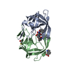
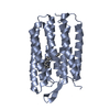
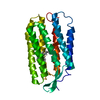
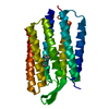
- PDB-4apv: The Klebsiella pneumoniae primosomal PriB protein: identification... -
+ Open data

- Basic information
| Entry | Database: PDB / ID: 4apv | ||||||
|---|---|---|---|---|---|---|---|
| Title | The Klebsiella pneumoniae primosomal PriB protein: identification, crystal structure, and ssDNA binding mode | ||||||
 Components Components | PRIMOSOMAL REPLICATION PROTEIN N | ||||||
 Keywords Keywords | REPLICATION / PRIB PRIMOSOME SSDNA BINDING | ||||||
| Function / homology | Nucleic acid-binding proteins / OB fold (Dihydrolipoamide Acetyltransferase, E2P) / Beta Barrel / Mainly Beta / :  Function and homology information Function and homology information | ||||||
| Biological species |  KLEBSIELLA PNEUMONIAE (bacteria) KLEBSIELLA PNEUMONIAE (bacteria) | ||||||
| Method |  X-RAY DIFFRACTION / SYNCHROTRON / MOLECULAR REPLACEMENT / Resolution: 2.095 Å X-RAY DIFFRACTION / SYNCHROTRON / MOLECULAR REPLACEMENT / Resolution: 2.095 Å | ||||||
 Authors Authors | Lo, Y.H. / Huang, Y.H. / Hsiao, C.D. / Huang, C.Y. | ||||||
 Citation Citation | Journal: Genes Cells / Year: 2012 Title: Crystal Structure and DNA-Binding Mode of Klebsiella Pneumoniae Primosomal Prib Protein. Authors: Huang, Y. / Lo, Y.H. / Huang, W. / Huang, C.Y. | ||||||
| History |
| ||||||
| Remark 700 | SHEET DETERMINATION METHOD: DSSP THE SHEETS PRESENTED AS "AA" IN EACH CHAIN ON SHEET RECORDS BELOW ... SHEET DETERMINATION METHOD: DSSP THE SHEETS PRESENTED AS "AA" IN EACH CHAIN ON SHEET RECORDS BELOW IS ACTUALLY AN 5-STRANDED BARREL THIS IS REPRESENTED BY A 6-STRANDED SHEET IN WHICH THE FIRST AND LAST STRANDS ARE IDENTICAL. |
- Structure visualization
| Structure viewer | Molecule: MolmilJmol/JSmol |
|---|
- Downloads & links
-Download
| PDBx/mmCIF format | 4apv.cif.gz | 51.8 KB | Display | PDBx/mmCIF format |
|---|---|---|---|---|
| PDB format | pdb4apv.ent.gz | 37.3 KB | Display | PDB format |
| PDBx/mmJSON format | 4apv.json.gz | Tree view | PDBx/mmJSON format | |
| Others |  Other downloads Other downloads |
-Validation report
| Arichive directory | https://data.pdbj.org/pub/pdb/validation_reports/ap/4apvftp://data.pdbj.org/pub/pdb/validation_reports/ap/4apv | HTTPS FTP |
|---|
-Related structure data
| Related structure data |  1v1qS S: Starting model for refinement |
|---|---|
| Similar structure data |
-Links
 PDBj
PDBj- Assembly
| Deposited unit | 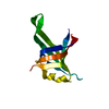
| ||||||||
|---|---|---|---|---|---|---|---|---|---|
| 1 | 
| ||||||||
| Unit cell |
|
-Components
| #1: Protein | Mass: 12559.387 Da / Num. of mol.: 1 Source method: isolated from a genetically manipulated source Source: (gene. exp.) KLEBSIELLA PNEUMONIAE (bacteria) / Production host: |
|---|---|
| #2: Water | ChemComp-HOH /  Mass: 18.015 Da / Num. of mol.: 10 / Source method: isolated from a natural source / Formula: H2O Mass: 18.015 Da / Num. of mol.: 10 / Source method: isolated from a natural source / Formula: H2O |
| Has protein modification | Y |
-Experimental details
-Experiment
| Experiment | Method: X-RAY DIFFRACTION / Number of used crystals: 10 |
|---|
- Sample preparation
| Crystal | Density Matthews: 1.85 Å3/Da / Density % sol: 33.4 % / Description: NONE |
|---|---|
| Crystal grow | pH: 5 / Details: 50% ETHANOL, 10 MM SODIUM ACETATE |
-Data collection
| Diffraction | Mean temperature: 298 K |
|---|---|
| Diffraction source | Source: SYNCHROTRON / Site: NSRRC  / Beamline: BL13B1 / Wavelength: 0.97622 / Beamline: BL13B1 / Wavelength: 0.97622 |
| Detector | Type: ADSC QUANTUM 315r / Detector: CCD / Date: Jan 20, 2010 |
| Radiation | Protocol: SINGLE WAVELENGTH / Monochromatic (M) / Laue (L): M / Scattering type: x-ray |
| Radiation wavelength | Wavelength: 0.97622 Å / Relative weight: 1 |
| Reflection | Resolution: 2.1→29.4 Å / Num. obs: 5031 / % possible obs: 95.6 % / Observed criterion σ(I): 2 / Redundancy: 3.4 % / Biso Wilson estimate: 34.96 Å2 / Rmerge(I) obs: 0.06 / Net I/σ(I): 40.7 |
| Reflection shell | Resolution: 2.09→2.1 Å / Redundancy: 3.6 % / Rmerge(I) obs: 0.26 / Mean I/σ(I) obs: 6.4 / % possible all: 99.8 |
- Processing
| Software |
| ||||||||||||||||||||||||||||||||||||||||||||||||||||||||||||||||||||||||||||||||||||||||||||||||||||||||||||||||||||||||||||||||||||||||||||||||||||||
|---|---|---|---|---|---|---|---|---|---|---|---|---|---|---|---|---|---|---|---|---|---|---|---|---|---|---|---|---|---|---|---|---|---|---|---|---|---|---|---|---|---|---|---|---|---|---|---|---|---|---|---|---|---|---|---|---|---|---|---|---|---|---|---|---|---|---|---|---|---|---|---|---|---|---|---|---|---|---|---|---|---|---|---|---|---|---|---|---|---|---|---|---|---|---|---|---|---|---|---|---|---|---|---|---|---|---|---|---|---|---|---|---|---|---|---|---|---|---|---|---|---|---|---|---|---|---|---|---|---|---|---|---|---|---|---|---|---|---|---|---|---|---|---|---|---|---|---|---|---|---|---|
| Refinement | Method to determine structure: MOLECULAR REPLACEMENT Starting model: PDB ENTRY 1V1Q Resolution: 2.095→26.649 Å / SU ML: 0.3 / σ(F): 1.37 / Phase error: 28.3 / Stereochemistry target values: ML
| ||||||||||||||||||||||||||||||||||||||||||||||||||||||||||||||||||||||||||||||||||||||||||||||||||||||||||||||||||||||||||||||||||||||||||||||||||||||
| Solvent computation | Shrinkage radii: 0.86 Å / VDW probe radii: 1.1 Å / Solvent model: FLAT BULK SOLVENT MODEL / Bsol: 66.888 Å2 / ksol: 0.389 e/Å3 | ||||||||||||||||||||||||||||||||||||||||||||||||||||||||||||||||||||||||||||||||||||||||||||||||||||||||||||||||||||||||||||||||||||||||||||||||||||||
| Displacement parameters | Biso mean: 52.9 Å2
| ||||||||||||||||||||||||||||||||||||||||||||||||||||||||||||||||||||||||||||||||||||||||||||||||||||||||||||||||||||||||||||||||||||||||||||||||||||||
| Refinement step | Cycle: LAST / Resolution: 2.095→26.649 Å
| ||||||||||||||||||||||||||||||||||||||||||||||||||||||||||||||||||||||||||||||||||||||||||||||||||||||||||||||||||||||||||||||||||||||||||||||||||||||
| Refine LS restraints |
| ||||||||||||||||||||||||||||||||||||||||||||||||||||||||||||||||||||||||||||||||||||||||||||||||||||||||||||||||||||||||||||||||||||||||||||||||||||||
| LS refinement shell |
| ||||||||||||||||||||||||||||||||||||||||||||||||||||||||||||||||||||||||||||||||||||||||||||||||||||||||||||||||||||||||||||||||||||||||||||||||||||||
| Refinement TLS params. | Method: refined / Refine-ID: X-RAY DIFFRACTION
| ||||||||||||||||||||||||||||||||||||||||||||||||||||||||||||||||||||||||||||||||||||||||||||||||||||||||||||||||||||||||||||||||||||||||||||||||||||||
| Refinement TLS group |
|
