Mass: 18.015 Da / Num. of mol.: 38 / Source method: isolated from a natural source / Formula: H2O
Has protein modification
Y
Sequence details
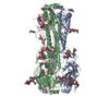
THROMBIN SITE WAS INTRODUCED BETWEEN RESIDUES 29 AND 30 OF EXOU. THE DIGESTED PROTEIN WAS ...THROMBIN SITE WAS INTRODUCED BETWEEN RESIDUES 29 AND 30 OF EXOU. THE DIGESTED PROTEIN WAS CRYSTALLIZED IN COMPLEX WITH SPCU
-
Experimental details
-
Experiment
Experiment
Method: X-RAY DIFFRACTION / Number of used crystals: 1
-
Sample preparation
Crystal
Density Matthews: 2.4 Å3/Da / Density % sol: 49 % / Description: NONE
Protocol: SINGLE WAVELENGTH / Monochromatic (M) / Laue (L): M / Scattering type: x-ray
Radiation wavelength
Wavelength: 0.97908 Å / Relative weight: 1
Reflection
Resolution: 2.94→97.05 Å / Num. obs: 17008 / % possible obs: 98.1 % / Observed criterion σ(I): 3 / Redundancy: 6.4 % / Biso Wilson estimate: 82.666 Å2 / Rmerge(I) obs: 0.09 / Net I/σ(I): 13.1
Reflection shell
Resolution: 2.94→3.12 Å / Redundancy: 6.3 % / Rmerge(I) obs: 0.67 / Mean I/σ(I) obs: 3 / % possible all: 91.6
-
Processing
Software
Name
Version
Classification
REFMAC
5.6.0117
refinement
XDS
datareduction
XSCALE
datascaling
PHENIX
phasing
Refinement
Method to determine structure: SAD Starting model: NONE Resolution: 2.94→97.05 Å / Cor.coef. Fo:Fc: 0.939 / Cor.coef. Fo:Fc free: 0.923 / SU B: 42.898 / SU ML: 0.378 / Cross valid method: THROUGHOUT / ESU R Free: 0.448 / Stereochemistry target values: MAXIMUM LIKELIHOOD Details: HYDROGENS HAVE BEEN ADDED IN THE RIDING POSITIONS. HYDROGENS HAVE BEEN USED IF PRESENT IN THE INPUT.
Rfactor
Num. reflection
% reflection
Selection details
Rfree
0.26437
840
5 %
RANDOM
Rwork
0.20801
-
-
-
obs
0.21102
15943
98.68 %
-
Solvent computation
Ion probe radii: 0.8 Å / Shrinkage radii: 0.8 Å / VDW probe radii: 1.2 Å / Solvent model: MASK
 Movie
Movie Controller

 Yorodumi
 Open data
 Basic information
 Components
 Keywords
 Function and homology information
 PSEUDOMONAS AERUGINOSA (bacteria)
 X-RAY DIFFRACTION /
  Authors
 Citation
 Structure visualization
 Downloads & links
 Other downloads


 PDBj
PDBj

 Assembly
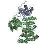
 Mass: 18.015 Da / Num. of mol.: 38 / Source method: isolated from a natural source / Formula: H2O
 Sample preparation
 / Beamline: ID29 / Wavelength: 0.97908
  Processing