 Movie
Movie Controller

[English] 日本語
 Yorodumi
- PDB-4ako: Mutations in the neighbourhood of CotA-laccase trinuclear site: E... -
+ Open data

- Basic information
| Entry | Database: PDB / ID: 4ako | ||||||
|---|---|---|---|---|---|---|---|
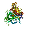
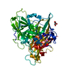
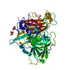
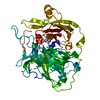
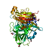
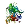
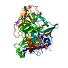
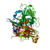
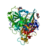
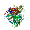
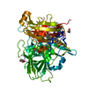
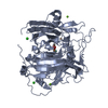
| Title | Mutations in the neighbourhood of CotA-laccase trinuclear site: E498L mutant | ||||||
 Components Components | SPORE COAT PROTEIN A | ||||||
 Keywords Keywords | OXIDOREDUCTASE / MULTI-COPPER OXIDASE / TRINUCLEAR CLUSTER / OXYGEN REDUCTION | ||||||
| Function / homology |  Function and homology information Function and homology informationbilirubin oxidase / bilirubin oxidase activity / hydroquinone:oxygen oxidoreductase activity / laccase / sporulation resulting in formation of a cellular spore / outer membrane-bounded periplasmic space / oxidoreductase activity / copper ion binding Similarity search - Function | ||||||
| Biological species |  | ||||||
| Method |  X-RAY DIFFRACTION / SYNCHROTRON / MOLECULAR REPLACEMENT / Resolution: 1.7 Å X-RAY DIFFRACTION / SYNCHROTRON / MOLECULAR REPLACEMENT / Resolution: 1.7 Å | ||||||
 Authors Authors | Silva, C.S. / Chen, Z. / Durao, P. / Pereira, M.M. / Todorovic, S. / Hildebrandt, P. / Martins, L.O. / Lindley, P.F. / Bento, I. | ||||||
 Citation Citation | Journal: Dalton Trans / Year: 2010 Title: The Role of Glu498 in the Dioxygen Reactivity of Cota-Laccase from Bacillus Subtilis. Authors: Chen, Z. / Durao, P. / Silva, C.S. / Pereira, M.M. / Todorovic, S. / Hildebrandt, P. / Bento, I. / Lindley, P.F. / Martins, L.O. | ||||||
| History |
|
- Structure visualization
| Structure viewer | Molecule: MolmilJmol/JSmol |
|---|
- Downloads & links
-Download
| PDBx/mmCIF format | 4ako.cif.gz | 133.2 KB | Display | PDBx/mmCIF format |
|---|---|---|---|---|
| PDB format | pdb4ako.ent.gz | 101.7 KB | Display | PDB format |
| PDBx/mmJSON format | 4ako.json.gz | Tree view | PDBx/mmJSON format | |
| Others |  Other downloads Other downloads |
-Validation report
| Arichive directory | https://data.pdbj.org/pub/pdb/validation_reports/ak/4akoftp://data.pdbj.org/pub/pdb/validation_reports/ak/4ako | HTTPS FTP |
|---|
-Related structure data
| Related structure data | 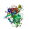 4akpC 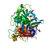 4akqC  1w6lS S: Starting model for refinement C: citing same article ( |
|---|---|
| Similar structure data |


-Links
 PDBj
PDBj


- Assembly
| Deposited unit | 
| ||||||||
|---|---|---|---|---|---|---|---|---|---|
| 1 |
| ||||||||
| Unit cell |
|
-Components
| #1: Protein | Mass: 58574.836 Da / Num. of mol.: 1 / Mutation: YES Source method: isolated from a genetically manipulated source Details: ETHYLENE GLYCOL (EDO) AS SOLVENT, OXIDIZED CYSTEINE AT POSITION 35 (CSX) Source: (gene. exp.) | ||||||||||||
|---|---|---|---|---|---|---|---|---|---|---|---|---|---|
| #2: Chemical | ChemComp-CU /   Mass: 63.546 Da / Num. of mol.: 4 / Source method: obtained synthetically / Formula: Cu Mass: 63.546 Da / Num. of mol.: 4 / Source method: obtained synthetically / Formula: Cu#3: Chemical | ChemComp-OXY / |   Mass: 31.999 Da / Num. of mol.: 1 / Source method: obtained synthetically / Formula: O2 Mass: 31.999 Da / Num. of mol.: 1 / Source method: obtained synthetically / Formula: O2#4: Chemical | ChemComp-EDO /   Mass: 62.068 Da / Num. of mol.: 20 / Source method: obtained synthetically / Formula: C2H6O2 Mass: 62.068 Da / Num. of mol.: 20 / Source method: obtained synthetically / Formula: C2H6O2#5: Water | ChemComp-HOH / |  Mass: 18.015 Da / Num. of mol.: 559 / Source method: isolated from a natural source / Formula: H2O Mass: 18.015 Da / Num. of mol.: 559 / Source method: isolated from a natural source / Formula: H2OCompound details | ENGINEERED | Nonpolymer details | S-OXY CYSTEINE (CSX): COEXISTS WITH CYSTEINE AT POSITION 35 | Sequence details | AT RESIDUE 35, CYSTEINE AND OXY-CYSTEINE COEXIST | |
-Experimental details
-Experiment
| Experiment | Method: X-RAY DIFFRACTION / Number of used crystals: 1 |
|---|
- Sample preparation
| Crystal | Density Matthews: 3.52 Å3/Da / Density % sol: 65 % / Description: NONE |
|---|---|
| Crystal grow | pH: 5.5 Details: 8% PEG 4K, 0.1M SODIUM CITRATE PH5.5, 30% ISOPROPANOL |
-Data collection
| Diffraction | Mean temperature: 100 K |
|---|---|
| Diffraction source | Source: SYNCHROTRON / Site: ESRF  / Beamline: ID14-1 / Wavelength: 0.934 / Beamline: ID14-1 / Wavelength: 0.934 |
| Detector | Type: ADSC QUANTUM 210 / Detector: CCD / Date: Apr 20, 2007 |
| Radiation | Protocol: SINGLE WAVELENGTH / Monochromatic (M) / Laue (L): M / Scattering type: x-ray |
| Radiation wavelength | Wavelength: 0.934 Å / Relative weight: 1 |
| Reflection | Resolution: 1.7→50.8 Å / Num. obs: 89089 / % possible obs: 99.5 % / Observed criterion σ(I): 2.4 / Redundancy: 5.5 % / Rmerge(I) obs: 0.05 |
| Reflection shell | Resolution: 1.7→1.79 Å / Redundancy: 4.6 % / Rmerge(I) obs: 0.32 / % possible all: 100 |
- Processing
| Software |
| ||||||||||||||||||||||||||||||||||||||||||||||||||||||||||||||||||||||||||||||||||||||||||||||||||||||||||||||||||||||||||||||||||||||||||||||||||||||||||||||||||||||||||||||||||||||
|---|---|---|---|---|---|---|---|---|---|---|---|---|---|---|---|---|---|---|---|---|---|---|---|---|---|---|---|---|---|---|---|---|---|---|---|---|---|---|---|---|---|---|---|---|---|---|---|---|---|---|---|---|---|---|---|---|---|---|---|---|---|---|---|---|---|---|---|---|---|---|---|---|---|---|---|---|---|---|---|---|---|---|---|---|---|---|---|---|---|---|---|---|---|---|---|---|---|---|---|---|---|---|---|---|---|---|---|---|---|---|---|---|---|---|---|---|---|---|---|---|---|---|---|---|---|---|---|---|---|---|---|---|---|---|---|---|---|---|---|---|---|---|---|---|---|---|---|---|---|---|---|---|---|---|---|---|---|---|---|---|---|---|---|---|---|---|---|---|---|---|---|---|---|---|---|---|---|---|---|---|---|---|---|
| Refinement | Method to determine structure: MOLECULAR REPLACEMENT Starting model: PDB ENTRY 1W6L Resolution: 1.7→40.72 Å / Cor.coef. Fo:Fc: 0.961 / Cor.coef. Fo:Fc free: 0.955 / SU B: 1.466 / SU ML: 0.049 / Cross valid method: THROUGHOUT / ESU R: 0.082 / ESU R Free: 0.08 / Stereochemistry target values: MAXIMUM LIKELIHOOD Details: HYDROGENS HAVE BEEN ADDED IN THE RIDING POSITIONS. SYSTEMATICALLY DISORDERED REGION BETWEEN RESIDUES 212 AND 215. LOOP REGION BETWEEN RESIDUES 89 AND 97 POORLY DEFINED.
| ||||||||||||||||||||||||||||||||||||||||||||||||||||||||||||||||||||||||||||||||||||||||||||||||||||||||||||||||||||||||||||||||||||||||||||||||||||||||||||||||||||||||||||||||||||||
| Solvent computation | Ion probe radii: 0.8 Å / Shrinkage radii: 0.8 Å / VDW probe radii: 1.2 Å / Solvent model: MASK | ||||||||||||||||||||||||||||||||||||||||||||||||||||||||||||||||||||||||||||||||||||||||||||||||||||||||||||||||||||||||||||||||||||||||||||||||||||||||||||||||||||||||||||||||||||||
| Displacement parameters | Biso mean: 19.474 Å2
| ||||||||||||||||||||||||||||||||||||||||||||||||||||||||||||||||||||||||||||||||||||||||||||||||||||||||||||||||||||||||||||||||||||||||||||||||||||||||||||||||||||||||||||||||||||||
| Refinement step | Cycle: LAST / Resolution: 1.7→40.72 Å
| ||||||||||||||||||||||||||||||||||||||||||||||||||||||||||||||||||||||||||||||||||||||||||||||||||||||||||||||||||||||||||||||||||||||||||||||||||||||||||||||||||||||||||||||||||||||
| Refine LS restraints |
|