 Movie
Movie Controller

[English] 日本語
 Yorodumi
- PDB-4akm: Crystal structure of the human lysosome-associated membrane prote... -
+ Open data

- Basic information
| Entry | Database: PDB / ID: 4akm | ||||||
|---|---|---|---|---|---|---|---|
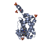
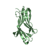
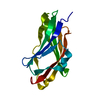
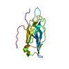
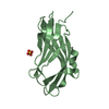
| Title | Crystal structure of the human lysosome-associated membrane protein LAMP-3 (aka DC-LAMP) | ||||||
 Components Components | LYSOSOME-ASSOCIATED MEMBRANE GLYCOPROTEIN 3 | ||||||
 Keywords Keywords | MEMBRANE PROTEIN / GLYCOSYLATION / BETA PRISM | ||||||
| Function / homology |  Function and homology information Function and homology informationregulation of viral life cycle / alveolar lamellar body membrane / establishment of protein localization to organelle / response to interferon-alpha / negative regulation of proteasomal protein catabolic process / regulation of autophagy / late endosome membrane / early endosome membrane / vesicle / adaptive immune response ...regulation of viral life cycle / alveolar lamellar body membrane / establishment of protein localization to organelle / response to interferon-alpha / negative regulation of proteasomal protein catabolic process / regulation of autophagy / late endosome membrane / early endosome membrane / vesicle / adaptive immune response / early endosome / lysosomal membrane / positive regulation of gene expression / perinuclear region of cytoplasm / cell surface / plasma membrane Similarity search - Function | ||||||
| Biological species |  HOMO SAPIENS (human) HOMO SAPIENS (human) | ||||||
| Method |  X-RAY DIFFRACTION / SYNCHROTRON / MAD / Resolution: 2.687 Å X-RAY DIFFRACTION / SYNCHROTRON / MAD / Resolution: 2.687 Å | ||||||
 Authors Authors | Krausze, J. / Wilke, S. / Buessow, K. | ||||||
 Citation Citation | Journal: Bmc Biol. / Year: 2012 Title: Crystal Structure of the Conserved Domain of the Dc Lysosomal Associated Membrane Protein: Implications for the Lysosomal Glycocalyx. Authors: Wilke, S. / Krausze, J. / Bussow, K. | ||||||
| History |
|
- Structure visualization
| Structure viewer | Molecule: MolmilJmol/JSmol |
|---|
- Downloads & links
-Download
| PDBx/mmCIF format | 4akm.cif.gz | 74.7 KB | Display | PDBx/mmCIF format |
|---|---|---|---|---|
| PDB format | pdb4akm.ent.gz | 56.1 KB | Display | PDB format |
| PDBx/mmJSON format | 4akm.json.gz | Tree view | PDBx/mmJSON format | |
| Others |  Other downloads Other downloads |
-Validation report
| Arichive directory | https://data.pdbj.org/pub/pdb/validation_reports/ak/4akmftp://data.pdbj.org/pub/pdb/validation_reports/ak/4akm | HTTPS FTP |
|---|
-Related structure data
| Similar structure data |
|---|


-Links
 PDBj
PDBj- Assembly
| Deposited unit | 
| ||||||||
|---|---|---|---|---|---|---|---|---|---|
| 1 | 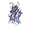
| ||||||||
| 2 | 
| ||||||||
| Unit cell |
| ||||||||
| Noncrystallographic symmetry (NCS) | NCS oper: (Code: given Matrix: (0.9291, 0.35759, 0.09437), Vector: |
-Components
| #1: Protein | Mass: 18656.707 Da / Num. of mol.: 2 Fragment: MEMBRANE-PROXIMAL PART OF LUMINAL DOMAIN, RESIDUES 222-381 Source method: isolated from a genetically manipulated source Source: (gene. exp.) HOMO SAPIENS (human) / Cell line (production host): CHO LEC3.2.8.1 / Production host:   CRICETULUS GRISEUS (Chinese hamster) / References: UniProt: Q9UQV4 CRICETULUS GRISEUS (Chinese hamster) / References: UniProt: Q9UQV4#2: Chemical | ChemComp-IR3 /   Mass: 192.217 Da / Num. of mol.: 4 / Source method: obtained synthetically / Formula: Ir Mass: 192.217 Da / Num. of mol.: 4 / Source method: obtained synthetically / Formula: Ir#3: Sugar |   Type: D-saccharide, beta linking / Mass: 221.208 Da / Num. of mol.: 2 Type: D-saccharide, beta linking / Mass: 221.208 Da / Num. of mol.: 2Source method: isolated from a genetically manipulated source Formula: C8H15NO6 #4: Water | ChemComp-HOH / |  Mass: 18.015 Da / Num. of mol.: 10 / Source method: isolated from a natural source / Formula: H2O Mass: 18.015 Da / Num. of mol.: 10 / Source method: isolated from a natural source / Formula: H2OHas protein modification | Y | Nonpolymer details | N-ACETYL-D-GLUCOSAMIN | |
|---|
-Experimental details
-Experiment
| Experiment | Method: X-RAY DIFFRACTION / Number of used crystals: 1 |
|---|
- Sample preparation
| Crystal | Density Matthews: 3.28 Å3/Da / Density % sol: 62.5 % / Description: NONE |
|---|---|
| Crystal grow | pH: 5 Details: PROTEIN WAS CRYSTALLIZED FROM 5% PEG 6000, 0.1 M CITRIC ACID, PH 5.0; THEN SOAKED IN 0.014 M AMMONIUM HEXACHLOROIRIDATE(III)HYDRATE AND CRYO-PROTECTED IN 30% PEG 6000. |
-Data collection
| Diffraction | Mean temperature: 100 K | ||||||||||||
|---|---|---|---|---|---|---|---|---|---|---|---|---|---|
| Diffraction source | Source: SYNCHROTRON / Site: EMBL/DESY, HAMBURG  / Beamline: X12 / Wavelength: 1.10371, 1.1042, 1.1009 / Beamline: X12 / Wavelength: 1.10371, 1.1042, 1.1009 | ||||||||||||
| Detector | Type: MARRESEARCH / Detector: CCD / Date: May 7, 2010 / Details: MIRROR | ||||||||||||
| Radiation | Monochromator: SI(111) / Protocol: MAD / Monochromatic (M) / Laue (L): M / Scattering type: x-ray | ||||||||||||
| Radiation wavelength |
| ||||||||||||
| Reflection | Resolution: 2.7→20 Å / Num. obs: 11238 / % possible obs: 91.1 % / Observed criterion σ(I): 2.5 / Redundancy: 2.86 % / Biso Wilson estimate: 55.49 Å2 / Rmerge(I) obs: 0.05 / Net I/σ(I): 15.48 | ||||||||||||
| Reflection shell | Resolution: 2.7→2.85 Å / Redundancy: 2.84 % / Rmerge(I) obs: 0.46 / Mean I/σ(I) obs: 2.47 / % possible all: 97.9 |
- Processing
| Software |
| |||||||||||||||||||||||||||||||||||
|---|---|---|---|---|---|---|---|---|---|---|---|---|---|---|---|---|---|---|---|---|---|---|---|---|---|---|---|---|---|---|---|---|---|---|---|---|
| Refinement | Method to determine structure: MAD Starting model: NONE Resolution: 2.687→19.295 Å / SU ML: 0.87 / σ(F): 1.98 / Phase error: 29.06 / Stereochemistry target values: MLHL
| |||||||||||||||||||||||||||||||||||
| Solvent computation | Shrinkage radii: 1.11 Å / VDW probe radii: 1.3 Å / Solvent model: FLAT BULK SOLVENT MODEL / Bsol: 10.238 Å2 / ksol: 0.235 e/Å3 | |||||||||||||||||||||||||||||||||||
| Displacement parameters | Biso mean: 61.3 Å2
| |||||||||||||||||||||||||||||||||||
| Refinement step | Cycle: LAST / Resolution: 2.687→19.295 Å
| |||||||||||||||||||||||||||||||||||
| Refine LS restraints |
| |||||||||||||||||||||||||||||||||||
| LS refinement shell |
|
