 Movie
Movie Controller

[English] 日本語
 Yorodumi
- PDB-4aiz: Crystallographic structure of 3mJL2 from the germinal line lambda 3 -
+ Open data

- Basic information
| Entry | Database: PDB / ID: 4aiz | ||||||
|---|---|---|---|---|---|---|---|
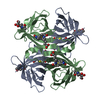
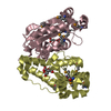
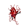
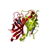
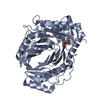
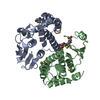
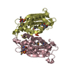
| Title | Crystallographic structure of 3mJL2 from the germinal line lambda 3 | ||||||
 Components Components | (V2-17 PROTEIN) x 2 | ||||||
 Keywords Keywords | IMMUNE SYSTEM / AMYLOIDOSIS | ||||||
| Function / homology |  Function and homology information Function and homology informationFc epsilon receptor (FCERI) signaling / Classical antibody-mediated complement activation / Initial triggering of complement / FCGR activation / Role of LAT2/NTAL/LAB on calcium mobilization / Role of phospholipids in phagocytosis / immunoglobulin complex / Scavenging of heme from plasma / FCERI mediated Ca+2 mobilization / FCGR3A-mediated IL10 synthesis ...Fc epsilon receptor (FCERI) signaling / Classical antibody-mediated complement activation / Initial triggering of complement / FCGR activation / Role of LAT2/NTAL/LAB on calcium mobilization / Role of phospholipids in phagocytosis / immunoglobulin complex / Scavenging of heme from plasma / FCERI mediated Ca+2 mobilization / FCGR3A-mediated IL10 synthesis / Regulation of Complement cascade / Cell surface interactions at the vascular wall / FCGR3A-mediated phagocytosis / FCERI mediated MAPK activation / Regulation of actin dynamics for phagocytic cup formation / Immunoregulatory interactions between a Lymphoid and a non-Lymphoid cell / FCERI mediated NF-kB activation / adaptive immune response / extracellular region / plasma membrane Similarity search - Function | ||||||
| Biological species |  HOMO SAPIENS (human) HOMO SAPIENS (human) | ||||||
| Method |  X-RAY DIFFRACTION / SYNCHROTRON / MOLECULAR REPLACEMENT / Resolution: 1.75 Å X-RAY DIFFRACTION / SYNCHROTRON / MOLECULAR REPLACEMENT / Resolution: 1.75 Å | ||||||
 Authors Authors | Villalba, M.I. / Luna, O.D. / Rudino-Pinera, E. / Sanchez, R. / Sanchez-Lopez, R. / Rojas-Trejo, S. / Olamendi-Portugal, T. / Fernandez-Velasco, D.A. / Becerril, B. | ||||||
 Citation Citation | Journal: J.Biol.Chem. / Year: 2015 Title: Site-Directed Mutagenesis Reveals Regions Implicated in the Stability and Fiber Formation of Human Lambda3R Light Chains. Authors: Villalba, M.I. / Canul-Tec, J.C. / Luna-Martinez, O.D. / Sanchez-Alcala, R. / Olamendi-Portugal, T. / Rudino-Pinera, E. / Rojas, S. / Sanchez-Lopez, R. / Fernandez-Velasco, D.A. / Becerril, B. | ||||||
| History |
|
- Structure visualization
| Structure viewer | Molecule: MolmilJmol/JSmol |
|---|
- Downloads & links
-Download
| PDBx/mmCIF format | 4aiz.cif.gz | 104.3 KB | Display | PDBx/mmCIF format |
|---|---|---|---|---|
| PDB format | pdb4aiz.ent.gz | 81.5 KB | Display | PDB format |
| PDBx/mmJSON format | 4aiz.json.gz | Tree view | PDBx/mmJSON format | |
| Others |  Other downloads Other downloads |
-Validation report
| Arichive directory | https://data.pdbj.org/pub/pdb/validation_reports/ai/4aizftp://data.pdbj.org/pub/pdb/validation_reports/ai/4aiz | HTTPS FTP |
|---|
-Related structure data
| Related structure data |  4aixC 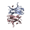 4aj0C  1lilS S: Starting model for refinement C: citing same article ( |
|---|---|
| Similar structure data |


-Links
 PDBj
PDBj


- Assembly
| Deposited unit | 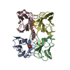
| ||||||||
|---|---|---|---|---|---|---|---|---|---|
| 1 |
| ||||||||
| Unit cell |
|
-Components
-Antibody , 2 types, 4 molecules ACDB
| #1: Antibody | Mass: 11517.674 Da / Num. of mol.: 3 Source method: isolated from a genetically manipulated source Source: (gene. exp.) HOMO SAPIENS (human) / Production host:  #2: Antibody | | Mass: 11515.701 Da / Num. of mol.: 1 Source method: isolated from a genetically manipulated source Source: (gene. exp.) HOMO SAPIENS (human) / Production host: |
|---|
-Non-polymers , 4 types, 409 molecules 


| #3: Chemical |  Mass: 96.063 Da / Num. of mol.: 2 / Source method: obtained synthetically / Formula: SO4 Mass: 96.063 Da / Num. of mol.: 2 / Source method: obtained synthetically / Formula: SO4#4: Chemical | ChemComp-CIT / |  Mass: 192.124 Da / Num. of mol.: 1 / Source method: obtained synthetically / Formula: C6H8O7 Mass: 192.124 Da / Num. of mol.: 1 / Source method: obtained synthetically / Formula: C6H8O7#5: Chemical | ChemComp-88Q / |  Mass: 262.299 Da / Num. of mol.: 1 / Source method: obtained synthetically / Formula: C12H22O6 Mass: 262.299 Da / Num. of mol.: 1 / Source method: obtained synthetically / Formula: C12H22O6#6: Water | ChemComp-HOH / | Mass: 18.015 Da / Num. of mol.: 405 / Source method: isolated from a natural source / Formula: H2O |
|---|
-Details
| Has protein modification | Y |
|---|---|
| Nonpolymer details | 88Q WAS CONSTRUCTED BASED ON THE ELECTRON DENSITY IN THE POSITION, HOWEVER WE WERE NOT ABLE TO FIND ...88Q WAS CONSTRUCTE |
| Sequence details | THIS DEPOSIT IS THE FIRST REFERENCE TO THE SEQUENCE OF 3MJL2. |
-Experimental details
-Experiment
| Experiment | Method: X-RAY DIFFRACTION / Number of used crystals: 1 |
|---|
- Sample preparation
| Crystal | Density Matthews: 2.1 Å3/Da / Density % sol: 41.33 % / Description: NONE |
|---|---|
| Crystal grow | pH: 7.5 Details: 0.2 M SODIUM CITRATE, 0.1 M HEPES, 30 % MPD, pH 7.5 |
-Data collection
| Diffraction | Mean temperature: 100 K |
|---|---|
| Diffraction source | Source: SYNCHROTRON / Site: NSLS  / Beamline: X6A / Wavelength: 0.933 / Beamline: X6A / Wavelength: 0.933 |
| Detector | Type: ADSC CCD / Detector: CCD / Date: May 1, 2011 Details: DOUBLE CRYSTAL CHANNEL CUT, SI(111), 1M LONG RH COATED TOROIDAL MIRROR FOR VERTICAL AND HORIZONTAL FOCUSING. |
| Radiation | Protocol: SINGLE WAVELENGTH / Monochromatic (M) / Laue (L): M / Scattering type: x-ray |
| Radiation wavelength | Wavelength: 0.933 Å / Relative weight: 1 |
| Reflection | Resolution: 1.75→30 Å / Num. obs: 31323 / % possible obs: 79.4 % / Observed criterion σ(I): 0 / Redundancy: 6.8 % / Biso Wilson estimate: 17.22 Å2 / Rmerge(I) obs: 0.07 / Net I/σ(I): 7.4 |
| Reflection shell | Resolution: 1.75→1.84 Å / Redundancy: 7.2 % / Rmerge(I) obs: 0.37 / Mean I/σ(I) obs: 2.1 / % possible all: 74.3 |
- Processing
| Software |
| ||||||||||||||||||||||||||||||||||||||||||||||||||||||||||||||||||||||||||||||||||||
|---|---|---|---|---|---|---|---|---|---|---|---|---|---|---|---|---|---|---|---|---|---|---|---|---|---|---|---|---|---|---|---|---|---|---|---|---|---|---|---|---|---|---|---|---|---|---|---|---|---|---|---|---|---|---|---|---|---|---|---|---|---|---|---|---|---|---|---|---|---|---|---|---|---|---|---|---|---|---|---|---|---|---|---|---|---|
| Refinement | Method to determine structure: MOLECULAR REPLACEMENT Starting model: PDB ENTRY 1LIL Resolution: 1.75→34.224 Å / SU ML: 0.38 / σ(F): 1.38 / Phase error: 23.31 / Stereochemistry target values: ML
| ||||||||||||||||||||||||||||||||||||||||||||||||||||||||||||||||||||||||||||||||||||
| Solvent computation | Shrinkage radii: 0.49 Å / VDW probe radii: 0.8 Å / Solvent model: FLAT BULK SOLVENT MODEL / Bsol: 61.115 Å2 / ksol: 0.404 e/Å3 | ||||||||||||||||||||||||||||||||||||||||||||||||||||||||||||||||||||||||||||||||||||
| Displacement parameters | Biso mean: 22.5 Å2
| ||||||||||||||||||||||||||||||||||||||||||||||||||||||||||||||||||||||||||||||||||||
| Refinement step | Cycle: LAST / Resolution: 1.75→34.224 Å
| ||||||||||||||||||||||||||||||||||||||||||||||||||||||||||||||||||||||||||||||||||||
| Refine LS restraints |
| ||||||||||||||||||||||||||||||||||||||||||||||||||||||||||||||||||||||||||||||||||||
| LS refinement shell |
|
