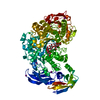
 Movie
Movie Controller

[English] 日本語
 Yorodumi
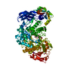
- PDB-4aio: Crystal structure of the starch debranching enzyme barley limit d... -
+ Open data

- Basic information
| Entry | Database: PDB / ID: 4aio | ||||||
|---|---|---|---|---|---|---|---|
| Title | Crystal structure of the starch debranching enzyme barley limit dextrinase | ||||||
 Components Components | LIMIT DEXTRINASE | ||||||
 Keywords Keywords | HYDROLASE / PULLULANASE / GLYCOSIDE HYDROLASE FAMILY 13 | ||||||
| Function / homology |  Function and homology information Function and homology informationpullulanase activity / polysaccharide catabolic process / metal ion binding Similarity search - Function | ||||||
| Biological species |  | ||||||
| Method |  X-RAY DIFFRACTION / SYNCHROTRON / MOLECULAR REPLACEMENT / Resolution: 1.9 Å X-RAY DIFFRACTION / SYNCHROTRON / MOLECULAR REPLACEMENT / Resolution: 1.9 Å | ||||||
 Authors Authors | Moeller, M.S. / Abou Hachem, M. / Svensson, B. / Henriksen, A. | ||||||
 Citation Citation | Journal: Acta Crystallogr.,Sect.F / Year: 2012 Title: Structure of the Starch-Debranching Enzyme Barley Limit Dextrinase Reveals Homology of the N-Terminal Domain to Cbm21. Authors: Moeller, M.S. / Abou Hachem, M. / Svensson, B. / Henriksen, A. #1: Journal: J.Mol.Biol. / Year: 2010Title: Crystal Structure of an Essential Enzyme in Seed Starch Degradation: Barley Limit Dextrinase in Complex with Cyclodextrins. Authors: Vester-Christensen, M.B. / Abou Hachem, M. / Svensson, B. / Henriksen, A. | ||||||
| History |
| ||||||
| Remark 700 | SHEET DETERMINATION METHOD: DSSP THE SHEETS PRESENTED AS "AA" IN EACH CHAIN ON SHEET RECORDS BELOW ... SHEET DETERMINATION METHOD: DSSP THE SHEETS PRESENTED AS "AA" IN EACH CHAIN ON SHEET RECORDS BELOW IS ACTUALLY AN 7-STRANDED BARREL THIS IS REPRESENTED BY A 8-STRANDED SHEET IN WHICH THE FIRST AND LAST STRANDS ARE IDENTICAL. SHEET DETERMINATION METHOD: DSSP THE SHEETS PRESENTED AS "AF" IN EACH CHAIN ON SHEET RECORDS BELOW IS ACTUALLY AN 8-STRANDED BARREL THIS IS REPRESENTED BY A 9-STRANDED SHEET IN WHICH THE FIRST AND LAST STRANDS ARE IDENTICAL. |
- Structure visualization
| Structure viewer | Molecule: MolmilJmol/JSmol |
|---|
- Downloads & links
-Download
| PDBx/mmCIF format | 4aio.cif.gz | 188.1 KB | Display | PDBx/mmCIF format |
|---|---|---|---|---|
| PDB format | pdb4aio.ent.gz | 146 KB | Display | PDB format |
| PDBx/mmJSON format | 4aio.json.gz | Tree view | PDBx/mmJSON format | |
| Others |  Other downloads Other downloads |
-Validation report
| Arichive directory | https://data.pdbj.org/pub/pdb/validation_reports/ai/4aioftp://data.pdbj.org/pub/pdb/validation_reports/ai/4aio | HTTPS FTP |
|---|
-Related structure data
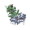
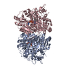
| Related structure data | 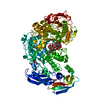 2y4sS S: Starting model for refinement |
|---|---|
| Similar structure data |


-Links
 PDBj
PDBj- Assembly
| Deposited unit | 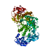
| ||||||||
|---|---|---|---|---|---|---|---|---|---|
| 1 |
| ||||||||
| Unit cell |
|
-Components
| #1: Protein | Mass: 97444.156 Da / Num. of mol.: 1 / Fragment: RESIDUES 22-904 / Mutation: YES Source method: isolated from a genetically manipulated source Source: (gene. exp.)  KOMAGATAELLA PASTORIS (fungus) / Strain (production host): GS115 / References: UniProt: O48541, pullulanase KOMAGATAELLA PASTORIS (fungus) / Strain (production host): GS115 / References: UniProt: O48541, pullulanase | ||||||||||
|---|---|---|---|---|---|---|---|---|---|---|---|
| #2: Chemical | ChemComp-GOL /   Mass: 92.094 Da / Num. of mol.: 4 / Source method: obtained synthetically / Formula: C3H8O3 Mass: 92.094 Da / Num. of mol.: 4 / Source method: obtained synthetically / Formula: C3H8O3#3: Chemical |   Mass: 40.078 Da / Num. of mol.: 2 / Source method: obtained synthetically / Formula: Ca Mass: 40.078 Da / Num. of mol.: 2 / Source method: obtained synthetically / Formula: Ca#4: Chemical | ChemComp-IOD /   Mass: 126.904 Da / Num. of mol.: 4 / Source method: obtained synthetically / Formula: I Mass: 126.904 Da / Num. of mol.: 4 / Source method: obtained synthetically / Formula: I#5: Water | ChemComp-HOH / |  Mass: 18.015 Da / Num. of mol.: 294 / Source method: isolated from a natural source / Formula: H2O Mass: 18.015 Da / Num. of mol.: 294 / Source method: isolated from a natural source / Formula: H2OCompound details | ENGINEERED | Sequence details | 4 AMINO ACID DISCREPANCIES. THE SEQUENCE STRETCH BETWEEN RESIDUES 484-486 REFLECTS THAT THE CLONED ...4 AMINO ACID DISCREPANC | |
-Experimental details
-Experiment
| Experiment | Method: X-RAY DIFFRACTION / Number of used crystals: 1 |
|---|
- Sample preparation
| Crystal | Density Matthews: 2.17 Å3/Da / Density % sol: 43.45 % / Description: NONE |
|---|---|
| Crystal grow | Details: PROTEIN STOCK: 10 MG/ML PROTEIN 50 MM MES PH 6.6, 250 MM NACL, 0.5 MM CACL2 AND 0.67 MM MALTOTRIOSE. RESERVOIR: 30% POLYETHYLENE GLYCOL (PEG) 3350, 5% GLYCEROL, AND 0.3 M NAI. CYSTEIN WAS ...Details: PROTEIN STOCK: 10 MG/ML PROTEIN 50 MM MES PH 6.6, 250 MM NACL, 0.5 MM CACL2 AND 0.67 MM MALTOTRIOSE. RESERVOIR: 30% POLYETHYLENE GLYCOL (PEG) 3350, 5% GLYCEROL, AND 0.3 M NAI. CYSTEIN WAS ADDED TO THE DROPS TO A FINAL CONCENTRATION OF 5-7 MM |
-Data collection
| Diffraction | Mean temperature: 100 K |
|---|---|
| Diffraction source | Source: SYNCHROTRON / Site: ESRF  / Beamline: ID23-1 / Wavelength: 0.97626 / Beamline: ID23-1 / Wavelength: 0.97626 |
| Detector | Type: ADSC QUANTUM 315r / Detector: CCD / Date: Nov 21, 2010 |
| Radiation | Protocol: SINGLE WAVELENGTH / Monochromatic (M) / Laue (L): M / Scattering type: x-ray |
| Radiation wavelength | Wavelength: 0.97626 Å / Relative weight: 1 |
| Reflection | Resolution: 1.9→35.54 Å / Num. obs: 60352 / % possible obs: 91.3 % / Redundancy: 2.6 % / Rmerge(I) obs: 0.04 / Net I/σ(I): 7.8 |
| Reflection shell | Resolution: 1.9→2 Å / Redundancy: 2.5 % / Rmerge(I) obs: 0.04 / Mean I/σ(I) obs: 2.1 / % possible all: 94.5 |
- Processing
| Software |
| ||||||||||||||||||||||||||||||||||||||||||||||||||||||||||||||||||||||||||||||||||||||||||||||||||||||||||||||||||||||||||||||||||||||||||||||||||||||||||||||||||||||||||||||||||||||
|---|---|---|---|---|---|---|---|---|---|---|---|---|---|---|---|---|---|---|---|---|---|---|---|---|---|---|---|---|---|---|---|---|---|---|---|---|---|---|---|---|---|---|---|---|---|---|---|---|---|---|---|---|---|---|---|---|---|---|---|---|---|---|---|---|---|---|---|---|---|---|---|---|---|---|---|---|---|---|---|---|---|---|---|---|---|---|---|---|---|---|---|---|---|---|---|---|---|---|---|---|---|---|---|---|---|---|---|---|---|---|---|---|---|---|---|---|---|---|---|---|---|---|---|---|---|---|---|---|---|---|---|---|---|---|---|---|---|---|---|---|---|---|---|---|---|---|---|---|---|---|---|---|---|---|---|---|---|---|---|---|---|---|---|---|---|---|---|---|---|---|---|---|---|---|---|---|---|---|---|---|---|---|---|
| Refinement | Method to determine structure: MOLECULAR REPLACEMENT Starting model: PDB ENTRY 2Y4S Resolution: 1.9→33.694 Å / Cor.coef. Fo:Fc: 0.947 / Cor.coef. Fo:Fc free: 0.923 / SU B: 3.322 / SU ML: 0.097 / Cross valid method: THROUGHOUT / ESU R: 0.185 / ESU R Free: 0.158 / Stereochemistry target values: MAXIMUM LIKELIHOOD / Details: HYDROGENS HAVE BEEN ADDED IN THE RIDING POSITIONS.
| ||||||||||||||||||||||||||||||||||||||||||||||||||||||||||||||||||||||||||||||||||||||||||||||||||||||||||||||||||||||||||||||||||||||||||||||||||||||||||||||||||||||||||||||||||||||
| Solvent computation | Ion probe radii: 0.8 Å / Shrinkage radii: 0.8 Å / VDW probe radii: 1.2 Å / Solvent model: MASK | ||||||||||||||||||||||||||||||||||||||||||||||||||||||||||||||||||||||||||||||||||||||||||||||||||||||||||||||||||||||||||||||||||||||||||||||||||||||||||||||||||||||||||||||||||||||
| Displacement parameters | Biso mean: 20.828 Å2
| ||||||||||||||||||||||||||||||||||||||||||||||||||||||||||||||||||||||||||||||||||||||||||||||||||||||||||||||||||||||||||||||||||||||||||||||||||||||||||||||||||||||||||||||||||||||
| Refinement step | Cycle: LAST / Resolution: 1.9→33.694 Å
| ||||||||||||||||||||||||||||||||||||||||||||||||||||||||||||||||||||||||||||||||||||||||||||||||||||||||||||||||||||||||||||||||||||||||||||||||||||||||||||||||||||||||||||||||||||||
| Refine LS restraints |
|