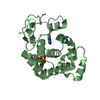
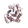
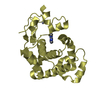
Entry Database : PDB / ID : 4ai4Title crystal structure of E38Q mutant of 3-methyladenine DNA glycosylase I from Staphylococcus aureus DNA-3-METHYLADENINE GLYCOSYLASE I Keywords / Function / homology / / / / Biological species STAPHYLOCOCCUS AUREUS SUBSP. AUREUS MSSA476 (bacteria)Method / / Resolution : 1.73 Å Authors Zhu, X. / Naismith, J.H. Journal : Acta Crystallogr.,Sect.F / Year : 2012Title : A Model for 3-Methyladenine Recognition by 3-Methyladenine DNA Glycosylase I (Tag) from Staphylococcus Aureus.Authors : Zhu, X. / Yan, X. / Carter, L.G. / Liu, H. / Graham, S. / Coote, P.J. / Naismith, J. History Deposition Feb 8, 2012 Deposition site / Processing site Revision 1.0 Feb 15, 2012 Provider / Type Revision 1.1 Jun 20, 2012 Group Revision 1.2 Apr 4, 2018 Group / Category / Item Revision 1.3 Dec 20, 2023 Group Data collection / Database references ... Data collection / Database references / Derived calculations / Other / Refinement description Category chem_comp_atom / chem_comp_bond ... chem_comp_atom / chem_comp_bond / database_2 / pdbx_database_status / pdbx_initial_refinement_model / pdbx_struct_conn_angle / struct_conn / struct_site Item _database_2.pdbx_DOI / _database_2.pdbx_database_accession ... _database_2.pdbx_DOI / _database_2.pdbx_database_accession / _pdbx_database_status.status_code_sf / _pdbx_struct_conn_angle.ptnr1_auth_comp_id / _pdbx_struct_conn_angle.ptnr1_auth_seq_id / _pdbx_struct_conn_angle.ptnr1_label_atom_id / _pdbx_struct_conn_angle.ptnr1_label_comp_id / _pdbx_struct_conn_angle.ptnr1_label_seq_id / _pdbx_struct_conn_angle.ptnr3_auth_comp_id / _pdbx_struct_conn_angle.ptnr3_auth_seq_id / _pdbx_struct_conn_angle.ptnr3_label_atom_id / _pdbx_struct_conn_angle.ptnr3_label_comp_id / _pdbx_struct_conn_angle.ptnr3_label_seq_id / _pdbx_struct_conn_angle.value / _struct_conn.pdbx_dist_value / _struct_conn.ptnr1_auth_comp_id / _struct_conn.ptnr1_auth_seq_id / _struct_conn.ptnr1_label_asym_id / _struct_conn.ptnr1_label_atom_id / _struct_conn.ptnr1_label_comp_id / _struct_conn.ptnr1_label_seq_id / _struct_conn.ptnr2_auth_comp_id / _struct_conn.ptnr2_auth_seq_id / _struct_conn.ptnr2_label_asym_id / _struct_conn.ptnr2_label_atom_id / _struct_conn.ptnr2_label_comp_id / _struct_conn.ptnr2_label_seq_id / _struct_site.pdbx_auth_asym_id / _struct_site.pdbx_auth_comp_id / _struct_site.pdbx_auth_seq_id
Show all Show less
 Movie
Movie Controller

 Yorodumi
 Open data
 Basic information
 Components
 Keywords
 Function and homology information
 STAPHYLOCOCCUS AUREUS SUBSP. AUREUS MSSA476 (bacteria)
 X-RAY DIFFRACTION /
  Authors
 Citation
 Structure visualization
 Downloads & links
 Other downloads


 PDBj
PDBj Assembly

 Mass: 65.409 Da / Num. of mol.: 1 / Source method: obtained synthetically / Formula: Zn
 Mass: 96.063 Da / Num. of mol.: 2 / Source method: obtained synthetically / Formula: SO4
 Mass: 18.015 Da / Num. of mol.: 164 / Source method: isolated from a natural source / Formula: H2O
 Sample preparation
 Processing