 Movie
Movie Controller

[English] 日本語
 Yorodumi
- PDB-4aha: Crystal Structure of Fucose binding lectin from Aspergillus Fumig... -
+ Open data

- Basic information
| Entry | Database: PDB / ID: 4aha | ||||||||||||
|---|---|---|---|---|---|---|---|---|---|---|---|---|---|
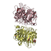
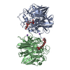
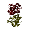
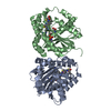
| Title | Crystal Structure of Fucose binding lectin from Aspergillus Fumigatus (AFL) in complex with fucosylated monosaccharides (Fuc1-2Gal, Fuc1- 3GlcNAc, Fuc1-4GlcNAc and Fuc1-6GlcNAc) | ||||||||||||
 Components Components | Fucose-specific lectin | ||||||||||||
 Keywords Keywords | SUGAR BINDING PROTEIN | ||||||||||||
| Function / homology |  Function and homology information Function and homology informationadhesion of symbiont to host cell surface via host glycoprotein / carbohydrate binding / metal ion binding Similarity search - Function | ||||||||||||
| Biological species |  | ||||||||||||
| Method |  X-RAY DIFFRACTION / SYNCHROTRON / MOLECULAR REPLACEMENT / Resolution: 2.2 Å X-RAY DIFFRACTION / SYNCHROTRON / MOLECULAR REPLACEMENT / Resolution: 2.2 Å | ||||||||||||
 Authors Authors | Houser, J. / Komarek, J. / Kostlanova, N. / Lahmann, M. / Cioci, G. / Varrot, A. / Imberty, A. / Wimmerova, M. | ||||||||||||
 Citation Citation | Journal: Acta Crystallogr.,Sect.D / Year: 2015 Title: Structural Insights Into Aspergillus Fumigatus Lectin Specificity: Afl Binding Sites are Functionally Non-Equivalent Authors: Houser, J. / Komarek, J. / Cioci, G. / Varrot, A. / Imberty, A. / Wimmerova, M. | ||||||||||||
| History |
|
- Structure visualization
| Structure viewer | Molecule: MolmilJmol/JSmol |
|---|
- Downloads & links
-Download
| PDBx/mmCIF format | 4aha.cif.gz | 147.9 KB | Display | PDBx/mmCIF format |
|---|---|---|---|---|
| PDB format | pdb4aha.ent.gz | 114.7 KB | Display | PDB format |
| PDBx/mmJSON format | 4aha.json.gz | Tree view | PDBx/mmJSON format | |
| Others |  Other downloads Other downloads |
-Validation report
| Arichive directory | https://data.pdbj.org/pub/pdb/validation_reports/ah/4ahaftp://data.pdbj.org/pub/pdb/validation_reports/ah/4aha | HTTPS FTP |
|---|
-Related structure data
| Related structure data | 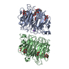 4agtC 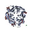 4ah4C 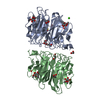 4c1yC 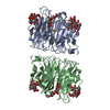 4d4uC  4d52C  4uouC  4agiS C: citing same article ( S: Starting model for refinement |
|---|---|
| Similar structure data |


-Links
 PDBj
PDBj
- Assembly
| Deposited unit | 
| ||||||||
|---|---|---|---|---|---|---|---|---|---|
| 1 |
| ||||||||
| Unit cell |
|
-Components
| #1: Protein | Mass: 34612.465 Da / Num. of mol.: 2 Source method: isolated from a genetically manipulated source Source: (gene. exp.)  #2: Polysaccharide | alpha-L-fucopyranose-(1-4)-2-acetamido-2-deoxy-beta-D-glucopyranose Source method: isolated from a genetically manipulated source #3: Sugar | ChemComp-FUC /   Type: L-saccharide, alpha linking / Mass: 164.156 Da / Num. of mol.: 8 Type: L-saccharide, alpha linking / Mass: 164.156 Da / Num. of mol.: 8Source method: isolated from a genetically manipulated source Formula: C6H12O5 #4: Water | ChemComp-HOH / |  Mass: 18.015 Da / Num. of mol.: 443 / Source method: isolated from a natural source / Formula: H2O Mass: 18.015 Da / Num. of mol.: 443 / Source method: isolated from a natural source / Formula: H2O |
|---|
-Experimental details
-Experiment
| Experiment | Method: X-RAY DIFFRACTION / Number of used crystals: 1 |
|---|
- Sample preparation
| Crystal | Density Matthews: 2.17 Å3/Da / Density % sol: 43.5 % / Description: NONE |
|---|---|
| Crystal grow | Details: 200 MM CACL2, 25% PEG 4K AND 100 MM TRIS, PH 8.5 |
-Data collection
| Diffraction | Mean temperature: 100 K |
|---|---|
| Diffraction source | Source: SYNCHROTRON / Site: ESRF  / Beamline: ID23-1 / Wavelength: 0.8726 / Beamline: ID23-1 / Wavelength: 0.8726 |
| Detector | Type: ADSC QUANTUM 315 / Detector: CCD / Details: MIRRORS |
| Radiation | Monochromator: SI111 / Protocol: SINGLE WAVELENGTH / Monochromatic (M) / Laue (L): M / Scattering type: x-ray |
| Radiation wavelength | Wavelength: 0.8726 Å / Relative weight: 1 |
| Reflection | Resolution: 2.2→36.41 Å / Num. obs: 27431 / % possible obs: 87.6 % / Observed criterion σ(I): 2 / Redundancy: 2.1 % / Biso Wilson estimate: 18.5 Å2 / Rmerge(I) obs: 0.12 / Net I/σ(I): 7.1 |
| Reflection shell | Resolution: 2.2→2.32 Å / Redundancy: 2.1 % / Rmerge(I) obs: 0.38 / Mean I/σ(I) obs: 2.7 / % possible all: 71.8 |
- Processing
| Software |
| ||||||||||||||||||||||||||||||||||||||||||||||||||||||||||||||||||||||||||||||||||||||||||||||||||||||||||||||||||||||||||||||||||||||||||||||||||||||||||||||||||||||||||||||||||||||
|---|---|---|---|---|---|---|---|---|---|---|---|---|---|---|---|---|---|---|---|---|---|---|---|---|---|---|---|---|---|---|---|---|---|---|---|---|---|---|---|---|---|---|---|---|---|---|---|---|---|---|---|---|---|---|---|---|---|---|---|---|---|---|---|---|---|---|---|---|---|---|---|---|---|---|---|---|---|---|---|---|---|---|---|---|---|---|---|---|---|---|---|---|---|---|---|---|---|---|---|---|---|---|---|---|---|---|---|---|---|---|---|---|---|---|---|---|---|---|---|---|---|---|---|---|---|---|---|---|---|---|---|---|---|---|---|---|---|---|---|---|---|---|---|---|---|---|---|---|---|---|---|---|---|---|---|---|---|---|---|---|---|---|---|---|---|---|---|---|---|---|---|---|---|---|---|---|---|---|---|---|---|---|---|
| Refinement | Method to determine structure: MOLECULAR REPLACEMENT Starting model: PDB ENTRY 4AGI Resolution: 2.2→77.47 Å / Cor.coef. Fo:Fc: 0.946 / Cor.coef. Fo:Fc free: 0.91 / SU B: 4.921 / SU ML: 0.125 / Cross valid method: THROUGHOUT / ESU R: 0.456 / ESU R Free: 0.228 / Stereochemistry target values: MAXIMUM LIKELIHOOD Details: HYDROGENS HAVE BEEN ADDED IN THE RIDING POSITIONS. U VALUES REFINED INDIVIDUALLY.
| ||||||||||||||||||||||||||||||||||||||||||||||||||||||||||||||||||||||||||||||||||||||||||||||||||||||||||||||||||||||||||||||||||||||||||||||||||||||||||||||||||||||||||||||||||||||
| Solvent computation | Ion probe radii: 0.8 Å / Shrinkage radii: 0.8 Å / VDW probe radii: 1.2 Å / Solvent model: MASK | ||||||||||||||||||||||||||||||||||||||||||||||||||||||||||||||||||||||||||||||||||||||||||||||||||||||||||||||||||||||||||||||||||||||||||||||||||||||||||||||||||||||||||||||||||||||
| Displacement parameters | Biso mean: 12.996 Å2
| ||||||||||||||||||||||||||||||||||||||||||||||||||||||||||||||||||||||||||||||||||||||||||||||||||||||||||||||||||||||||||||||||||||||||||||||||||||||||||||||||||||||||||||||||||||||
| Refinement step | Cycle: LAST / Resolution: 2.2→77.47 Å
| ||||||||||||||||||||||||||||||||||||||||||||||||||||||||||||||||||||||||||||||||||||||||||||||||||||||||||||||||||||||||||||||||||||||||||||||||||||||||||||||||||||||||||||||||||||||
| Refine LS restraints |
|