 Movie
Movie Controller


+ Open data

- Basic information
| Entry | Database: PDB / ID: 43fx | |||||||||
|---|---|---|---|---|---|---|---|---|---|---|
| Title | DPR epimerase complex, quabodepistat-bound state | |||||||||
 Components Components |
| |||||||||
 Keywords Keywords | OXIDOREDUCTASE / Epimerase complex | |||||||||
| Function / homology |  Function and homology information Function and homology informationdecaprenylphospho-beta-D-erythro-pentofuranosid-2-ulose 2-reductase / arabinan biosynthetic process / cell wall polysaccharide biosynthetic process / decaprenylphospho-beta-D-ribofuranose 2-dehydrogenase / D-arabinono-1,4-lactone oxidase activity / capsule polysaccharide biosynthetic process / FAD binding / cell wall organization / periplasmic space / oxidoreductase activity ...decaprenylphospho-beta-D-erythro-pentofuranosid-2-ulose 2-reductase / arabinan biosynthetic process / cell wall polysaccharide biosynthetic process / decaprenylphospho-beta-D-ribofuranose 2-dehydrogenase / D-arabinono-1,4-lactone oxidase activity / capsule polysaccharide biosynthetic process / FAD binding / cell wall organization / periplasmic space / oxidoreductase activity / response to antibiotic / plasma membrane Similarity search - Function | |||||||||
| Biological species |  Mycobacterium tuberculosis H37Rv (bacteria) Mycobacterium tuberculosis H37Rv (bacteria) | |||||||||
| Method | ELECTRON MICROSCOPY / single particle reconstruction / cryo EM / Resolution: 2.69 Å | |||||||||
 Authors Authors | Wu, F. / Gao, S. / Zhang, L. | |||||||||
| Funding support |  China, 1items China, 1items
| |||||||||
 Citation Citation | Journal: To Be Published Title: DPR epimerise complex, quabodepistat-bound state Authors: Wu, F. / Gao, S. / Zhang, L. | |||||||||
| History |
|
- Structure visualization
| Structure viewer | Molecule: MolmilJmol/JSmol |
|---|
- Downloads & links
-Download
| PDBx/mmCIF format | 43fx.cif.gz | 275.5 KB | Display | PDBx/mmCIF format |
|---|---|---|---|---|
| PDB format | pdb43fx.ent.gz | Display | PDB format | |
| PDBx/mmJSON format | 43fx.json.gz | Tree view | PDBx/mmJSON format | |
| Others |  Other downloads Other downloads |
-Validation report
| Arichive directory | https://data.pdbj.org/pub/pdb/validation_reports/3f/43fxftp://data.pdbj.org/pub/pdb/validation_reports/3f/43fx | HTTPS FTP |
|---|
-Related structure data
| Related structure data |  81853MC  62280 M: map data used to model this data C: citing same article ( |
|---|---|
| Similar structure data |
-Links
 PDBj
PDBj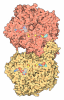
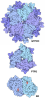
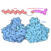
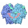
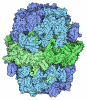
- Assembly
| Deposited unit | 
|
|---|---|
| 1 |
|
-Components
| #1: Protein | Mass: 50219.977 Da / Num. of mol.: 2 Source method: isolated from a genetically manipulated source Source: (gene. exp.) Mycobacterium tuberculosis H37Rv (bacteria)Gene: dprE1, Rv3790 Production host: Mycolicibacterium smegmatis MC2 155 (bacteria)References: UniProt: P9WJF1, decaprenylphospho-beta-D-ribofuranose 2-dehydrogenase #2: Protein | Mass: 27501.812 Da / Num. of mol.: 2 Source method: isolated from a genetically manipulated source Source: (gene. exp.) Mycobacterium tuberculosis H37Rv (bacteria)Gene: dprE2, Rv3791, MTCY13D12.25 Production host: Mycolicibacterium smegmatis MC2 155 (bacteria)References: UniProt: P9WGS9, decaprenylphospho-beta-D-erythro-pentofuranosid-2-ulose 2-reductase #3: Chemical |  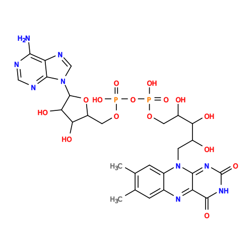 Mass: 785.550 Da / Num. of mol.: 2 / Source method: obtained synthetically / Formula: C27H33N9O15P2 / Feature type: SUBJECT OF INVESTIGATION / Comment: FAD*YM Mass: 785.550 Da / Num. of mol.: 2 / Source method: obtained synthetically / Formula: C27H33N9O15P2 / Feature type: SUBJECT OF INVESTIGATION / Comment: FAD*YM#4: Chemical | Mass: 456.843 Da / Num. of mol.: 2 / Source method: obtained synthetically / Formula: C21H20ClF3N2O4 / Feature type: SUBJECT OF INVESTIGATION Has ligand of interest | Y | Has protein modification | N | |
|---|
-Experimental details
-Experiment
| Experiment | Method: ELECTRON MICROSCOPY |
|---|---|
| EM experiment | Aggregation state: PARTICLE / 3D reconstruction method: single particle reconstruction |
- Sample preparation
| Component | Name: DPR epimerase complex / Type: COMPLEX / Entity ID: #1-#2 / Source: RECOMBINANT |
|---|---|
| Molecular weight | Value: 0.155 MDa / Experimental value: YES |
| Source (natural) | Organism: Mycobacterium tuberculosis H37Rv (bacteria) |
| Source (recombinant) | Organism: Mycolicibacterium smegmatis MC2 155 (bacteria) |
| Buffer solution | pH: 7.5 / Details: 150mM NaCl, 20mM Hepes |
| Specimen | Conc.: 0.5 mg/ml / Embedding applied: NO / Shadowing applied: NO / Staining applied: NO / Vitrification applied: YES |
| Specimen support | Grid material: COPPER / Grid mesh size: 300 divisions/in. / Grid type: Quantifoil R1.2/1.3 |
| Vitrification | Instrument: FEI VITROBOT MARK II / Cryogen name: ETHANE / Humidity: 100 % / Chamber temperature: 201 K |
- Electron microscopy imaging
| Experimental equipment |  Model: Talos Arctica / Image courtesy: FEI Company |
|---|---|
| Microscopy | Model: FEI TALOS ARCTICA |
| Electron gun | Electron source:  FIELD EMISSION GUN / Accelerating voltage: 300 kV / Illumination mode: SPOT SCAN FIELD EMISSION GUN / Accelerating voltage: 300 kV / Illumination mode: SPOT SCAN |
| Electron lens | Mode: BRIGHT FIELD / Nominal magnification: 105000 X / Nominal defocus max: 2000 nm / Nominal defocus min: 1200 nm / Alignment procedure: BASIC |
| Specimen holder | Cryogen: NITROGEN / Specimen holder model: FEI TITAN KRIOS AUTOGRID HOLDER |
| Image recording | Electron dose: 60 e/Å2 / Film or detector model: GATAN K3 BIOQUANTUM (6k x 4k) |
| EM imaging optics | Energyfilter name: GIF Bioquantum |
- Processing
| EM software |
| ||||||||||||||||||||||||
|---|---|---|---|---|---|---|---|---|---|---|---|---|---|---|---|---|---|---|---|---|---|---|---|---|---|
| CTF correction | Type: PHASE FLIPPING AND AMPLITUDE CORRECTION | ||||||||||||||||||||||||
| Symmetry | Point symmetry: C2 (2 fold cyclic) | ||||||||||||||||||||||||
| 3D reconstruction | Resolution: 2.69 Å / Resolution method: FSC 0.143 CUT-OFF / Num. of particles: 474208 / Algorithm: FOURIER SPACE / Num. of class averages: 1 / Symmetry type: POINT | ||||||||||||||||||||||||
| Refinement | Highest resolution: 2.69 Å Stereochemistry target values: REAL-SPACE (WEIGHTED MAP SUM AT ATOM CENTERS) | ||||||||||||||||||||||||
| Refine LS restraints |
|
