 Movie
Movie Controller

[English] 日本語
 Yorodumi
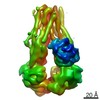
- PDB-3zef: Crystal structure of Prp8:Aar2 complex: second crystal form at 3.... -
+ Open data

- Basic information
| Entry | Database: PDB / ID: 3zef | ||||||
|---|---|---|---|---|---|---|---|
| Title | Crystal structure of Prp8:Aar2 complex: second crystal form at 3.1 Angstrom resolution | ||||||
 Components Components |
| ||||||
 Keywords Keywords | TRANSLATION / PRE-MRNA SPLICING / SPLICEOSOME / U5 SNRNP | ||||||
| Function / homology |  Function and homology information Function and homology informationgeneration of catalytic spliceosome for second transesterification step / mRNA 5'-splice site recognition / mRNA 3'-splice site recognition / spliceosomal tri-snRNP complex assembly / Prp19 complex / U5 snRNA binding / U5 snRNP / U2 snRNA binding / U6 snRNA binding / pre-mRNA intronic binding ...generation of catalytic spliceosome for second transesterification step / mRNA 5'-splice site recognition / mRNA 3'-splice site recognition / spliceosomal tri-snRNP complex assembly / Prp19 complex / U5 snRNA binding / U5 snRNP / U2 snRNA binding / U6 snRNA binding / pre-mRNA intronic binding / spliceosomal snRNP assembly / U1 snRNA binding / U4/U6 x U5 tri-snRNP complex / catalytic step 2 spliceosome / spliceosomal complex / mRNA splicing, via spliceosome / metallopeptidase activity / mRNA binding / nucleus / cytoplasm Similarity search - Function | ||||||
| Biological species |  | ||||||
| Method |  X-RAY DIFFRACTION / SYNCHROTRON / MOLECULAR REPLACEMENT / Resolution: 3.1 Å X-RAY DIFFRACTION / SYNCHROTRON / MOLECULAR REPLACEMENT / Resolution: 3.1 Å | ||||||
 Authors Authors | Galej, W.P. / Oubridge, C. / Newman, A.J. / Nagai, K. | ||||||
 Citation Citation | Journal: Nature / Year: 2013 Title: Crystal Structure of Prp8 Reveals Active Site Cavity of the Spliceosome Authors: Galej, W.P. / Oubridge, C. / Newman, A.J. / Nagai, K. | ||||||
| History |
|
- Structure visualization
| Structure viewer | Molecule: MolmilJmol/JSmol |
|---|
- Downloads & links
-Download
| PDBx/mmCIF format | 3zef.cif.gz | 714.6 KB | Display | PDBx/mmCIF format |
|---|---|---|---|---|
| PDB format | pdb3zef.ent.gz | 575.8 KB | Display | PDB format |
| PDBx/mmJSON format | 3zef.json.gz | Tree view | PDBx/mmJSON format | |
| Others |  Other downloads Other downloads |
-Validation report
| Summary document | 3zef_validation.pdf.gz | 492.9 KB | Display | wwPDB validaton report |
|---|---|---|---|---|
| Full document | 3zef_full_validation.pdf.gz | 653.6 KB | Display | |
| Data in XML | 3zef_validation.xml.gz | 135.2 KB | Display | |
| Data in CIF | 3zef_validation.cif.gz | 178.8 KB | Display | |
| Arichive directory | https://data.pdbj.org/pub/pdb/validation_reports/ze/3zefftp://data.pdbj.org/pub/pdb/validation_reports/ze/3zef | HTTPS FTP |
-Related structure data
| Related structure data | 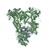 4i43C 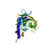 2og4S 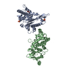 3sbtS C: citing same article ( S: Starting model for refinement |
|---|---|
| Similar structure data |


-Links
 PDBj
PDBj

- Assembly
| Deposited unit | 
| ||||||||
|---|---|---|---|---|---|---|---|---|---|
| 1 | 
| ||||||||
| 2 | 
| ||||||||
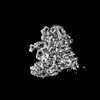
| Unit cell |
|
-Components
| #1: Protein | Mass: 41734.688 Da / Num. of mol.: 2 / Mutation: YES Source method: isolated from a genetically manipulated source Source: (gene. exp.) Plasmid: PRS424 / Production host: #2: Protein | Mass: 176632.312 Da / Num. of mol.: 2 Source method: isolated from a genetically manipulated source Source: (gene. exp.) Plasmid: PRS426 / Production host: Sequence details | THE DEPOSITED SEQUENCE REPRESENTS RESIDUES 885-2414 OF THE GENBANK DATABASE ENTRY WITH MUTATIONS ...THE DEPOSITED SEQUENCE REPRESENTS | |
|---|
-Experimental details
-Experiment
| Experiment | Method: X-RAY DIFFRACTION / Number of used crystals: 1 |
|---|
- Sample preparation
| Crystal | Density Matthews: 2.75 Å3/Da / Density % sol: 0.55 % Description: 3SBT SEPARATED INTO SINGLE CHAINS FOR MR SEARCH |
|---|---|
| Crystal grow | pH: 7.9 Details: 7-9% PEG 8000, 100 MM NA-CITRATE PH 7.9, 50-200 MM AMMONIUM SULFATE |
-Data collection
| Diffraction | Mean temperature: 100 K |
|---|---|
| Diffraction source | Source: SYNCHROTRON / Site: Diamond  / Beamline: I03 / Wavelength: 0.9795 / Beamline: I03 / Wavelength: 0.9795 |
| Detector | Type: DECTRIS PILATUS 6M / Detector: PIXEL / Date: Feb 6, 2012 |
| Radiation | Protocol: SINGLE WAVELENGTH / Monochromatic (M) / Laue (L): M / Scattering type: x-ray |
| Radiation wavelength | Wavelength: 0.9795 Å / Relative weight: 1 |
| Reflection | Resolution: 3.1→137.5 Å / Num. obs: 97506 / % possible obs: 99.9 % / Observed criterion σ(I): 2 / Redundancy: 5 % / Rmerge(I) obs: 0.16 / Net I/σ(I): 6.4 |
| Reflection shell | Resolution: 3.1→3.15 Å / Redundancy: 5.2 % / Rmerge(I) obs: 1.3 / Mean I/σ(I) obs: 1.4 / % possible all: 100 |
- Processing
| Software |
| ||||||||||||||||||||||||||||||||||||||||||||||||||||||||||||||||||||||||||||||||||||||||||||||||||||||||||||||||||||||||||||||||||||||||||||||||||||||||||||||||||||||||||||||||||||||
|---|---|---|---|---|---|---|---|---|---|---|---|---|---|---|---|---|---|---|---|---|---|---|---|---|---|---|---|---|---|---|---|---|---|---|---|---|---|---|---|---|---|---|---|---|---|---|---|---|---|---|---|---|---|---|---|---|---|---|---|---|---|---|---|---|---|---|---|---|---|---|---|---|---|---|---|---|---|---|---|---|---|---|---|---|---|---|---|---|---|---|---|---|---|---|---|---|---|---|---|---|---|---|---|---|---|---|---|---|---|---|---|---|---|---|---|---|---|---|---|---|---|---|---|---|---|---|---|---|---|---|---|---|---|---|---|---|---|---|---|---|---|---|---|---|---|---|---|---|---|---|---|---|---|---|---|---|---|---|---|---|---|---|---|---|---|---|---|---|---|---|---|---|---|---|---|---|---|---|---|---|---|---|---|
| Refinement | Method to determine structure: MOLECULAR REPLACEMENT Starting model: PDB ENTRIES 3SBT, 2OG4 Resolution: 3.1→137.45 Å / Cor.coef. Fo:Fc: 0.926 / Cor.coef. Fo:Fc free: 0.886 / SU B: 25.665 / SU ML: 0.441 / Cross valid method: THROUGHOUT / ESU R Free: 0.489 / Stereochemistry target values: MAXIMUM LIKELIHOOD Details: HYDROGENS HAVE BEEN ADDED IN THE RIDING POSITIONS. U VALUES ARE REFINED INDIVIDUALLY
| ||||||||||||||||||||||||||||||||||||||||||||||||||||||||||||||||||||||||||||||||||||||||||||||||||||||||||||||||||||||||||||||||||||||||||||||||||||||||||||||||||||||||||||||||||||||
| Solvent computation | Ion probe radii: 0.8 Å / Shrinkage radii: 0.8 Å / VDW probe radii: 1.2 Å / Solvent model: MASK | ||||||||||||||||||||||||||||||||||||||||||||||||||||||||||||||||||||||||||||||||||||||||||||||||||||||||||||||||||||||||||||||||||||||||||||||||||||||||||||||||||||||||||||||||||||||
| Displacement parameters | Biso mean: 95.693 Å2
| ||||||||||||||||||||||||||||||||||||||||||||||||||||||||||||||||||||||||||||||||||||||||||||||||||||||||||||||||||||||||||||||||||||||||||||||||||||||||||||||||||||||||||||||||||||||
| Refinement step | Cycle: LAST / Resolution: 3.1→137.45 Å
| ||||||||||||||||||||||||||||||||||||||||||||||||||||||||||||||||||||||||||||||||||||||||||||||||||||||||||||||||||||||||||||||||||||||||||||||||||||||||||||||||||||||||||||||||||||||
| Refine LS restraints |
|