 Movie
Movie Controller

[English] 日本語
 Yorodumi
- PDB-3zbm: Structure of M92A variant of three-domain heme-Cu nitrite reducta... -
+ Open data

- Basic information
| Entry | Database: PDB / ID: 3zbm | |||||||||
|---|---|---|---|---|---|---|---|---|---|---|
| Title | Structure of M92A variant of three-domain heme-Cu nitrite reductase from Ralstonia pickettii | |||||||||
 Components Components | COPPER-CONTAINING NITRITE REDUCTASE | |||||||||
 Keywords Keywords | OXIDOREDUCTASE / ELECTRON TRANSFER / PROTON CHANNEL / DENITRIFICATION | |||||||||
| Function / homology |  Function and homology information Function and homology informationnitrite reductase (NO-forming) / nitrite reductase (NO-forming) activity / periplasmic space / electron transfer activity / copper ion binding / heme binding Similarity search - Function | |||||||||
| Biological species |  RALSTONIA PICKETTII (bacteria) RALSTONIA PICKETTII (bacteria) | |||||||||
| Method |  X-RAY DIFFRACTION / SYNCHROTRON / FOURIER SYNTHESIS / Resolution: 1.87 Å X-RAY DIFFRACTION / SYNCHROTRON / FOURIER SYNTHESIS / Resolution: 1.87 Å | |||||||||
 Authors Authors | Antonyuk, S.V. / Han, C. / Eady, R.R. / Hasnain, S.S. | |||||||||
 Citation Citation | Journal: Nature / Year: 2013 Title: Structures of protein-protein complexes involved in electron transfer. Authors: Antonyuk, S.V. / Han, C. / Eady, R.R. / Hasnain, S.S. | |||||||||
| History |
|
- Structure visualization
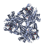
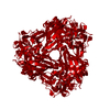
| Structure viewer | Molecule: MolmilJmol/JSmol |
|---|
- Downloads & links
-Download
| PDBx/mmCIF format | 3zbm.cif.gz | 115.4 KB | Display | PDBx/mmCIF format |
|---|---|---|---|---|
| PDB format | pdb3zbm.ent.gz | 88.5 KB | Display | PDB format |
| PDBx/mmJSON format | 3zbm.json.gz | Tree view | PDBx/mmJSON format | |
| Others |  Other downloads Other downloads |
-Validation report
| Arichive directory | https://data.pdbj.org/pub/pdb/validation_reports/zb/3zbmftp://data.pdbj.org/pub/pdb/validation_reports/zb/3zbm | HTTPS FTP |
|---|
-Related structure data
| Related structure data |  2yqbC 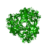 3ziyC 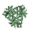 4ax3C  4aww C: citing same article ( S: Starting model for refinement |
|---|---|
| Similar structure data |


-Links
 PDBj
PDBj


- Assembly
| Deposited unit | 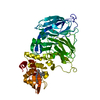
| |||||||||||||||
|---|---|---|---|---|---|---|---|---|---|---|---|---|---|---|---|---|
| 1 | 
| |||||||||||||||
| Unit cell |
| |||||||||||||||
| Components on special symmetry positions |
|
-Components
| #1: Protein | Mass: 49888.395 Da / Num. of mol.: 1 Source method: isolated from a genetically manipulated source Source: (gene. exp.) RALSTONIA PICKETTII (bacteria) / Strain: 12J / Description: (GENBANK ACCESSION NUMBER. NC_010678 / Plasmid: PET26B-RPNIR / Production host: References: UniProt: E2STD2, UniProt: B2UHR8*PLUS, nitrite reductase (NO-forming) | ||||||
|---|---|---|---|---|---|---|---|
| #2: Chemical |   Mass: 63.546 Da / Num. of mol.: 2 / Source method: obtained synthetically / Formula: Cu Mass: 63.546 Da / Num. of mol.: 2 / Source method: obtained synthetically / Formula: Cu#3: Chemical | ChemComp-HEC / |   Mass: 618.503 Da / Num. of mol.: 1 / Source method: obtained synthetically / Formula: C34H34FeN4O4 Mass: 618.503 Da / Num. of mol.: 1 / Source method: obtained synthetically / Formula: C34H34FeN4O4#4: Water | ChemComp-HOH / |  Mass: 18.015 Da / Num. of mol.: 457 / Source method: isolated from a natural source / Formula: H2O Mass: 18.015 Da / Num. of mol.: 457 / Source method: isolated from a natural source / Formula: H2OHas protein modification | Y | |
-Experimental details
-Experiment
| Experiment | Method: X-RAY DIFFRACTION / Number of used crystals: 1 |
|---|
- Sample preparation
| Crystal | Density Matthews: 2.78 Å3/Da / Density % sol: 56 % / Description: NONE |
|---|---|
| Crystal grow | pH: 6.5 Details: 20MM MES PH 6.5 20% PEG3350, 0.2 SODIUM CITRATE, 200 MM NACL, PROTEIN CONCENTRATION 7.5 MG/ML |
-Data collection
| Diffraction | Mean temperature: 100 K |
|---|---|
| Diffraction source | Source: SYNCHROTRON / Site: Diamond  / Beamline: I02 / Wavelength: 1 / Beamline: I02 / Wavelength: 1 |
| Detector | Type: DECTRIS PILATUS 6M / Detector: PIXEL / Date: Oct 11, 2012 / Details: MIRRORS |
| Radiation | Monochromator: SI111 / Protocol: SINGLE WAVELENGTH / Monochromatic (M) / Laue (L): M / Scattering type: x-ray |
| Radiation wavelength | Wavelength: 1 Å / Relative weight: 1 |
| Reflection | Resolution: 1.87→32 Å / Num. obs: 43074 / % possible obs: 98.9 % / Redundancy: 5 % / Biso Wilson estimate: 22.6 Å2 / Rmerge(I) obs: 0.12 / Net I/σ(I): 7.1 |
| Reflection shell | Resolution: 1.87→1.92 Å / Redundancy: 3 % / Rmerge(I) obs: 0.76 / Mean I/σ(I) obs: 1.8 / % possible all: 88.8 |
- Processing
| Software |
| ||||||||||||||||||||||||||||||||||||||||||||||||||||||||||||||||||||||||||||||||||||||||||||||||||||||||||||||||||||||||||||||||||||||||||||||||||||||||||||||||||||||||||||||||||||||
|---|---|---|---|---|---|---|---|---|---|---|---|---|---|---|---|---|---|---|---|---|---|---|---|---|---|---|---|---|---|---|---|---|---|---|---|---|---|---|---|---|---|---|---|---|---|---|---|---|---|---|---|---|---|---|---|---|---|---|---|---|---|---|---|---|---|---|---|---|---|---|---|---|---|---|---|---|---|---|---|---|---|---|---|---|---|---|---|---|---|---|---|---|---|---|---|---|---|---|---|---|---|---|---|---|---|---|---|---|---|---|---|---|---|---|---|---|---|---|---|---|---|---|---|---|---|---|---|---|---|---|---|---|---|---|---|---|---|---|---|---|---|---|---|---|---|---|---|---|---|---|---|---|---|---|---|---|---|---|---|---|---|---|---|---|---|---|---|---|---|---|---|---|---|---|---|---|---|---|---|---|---|---|---|
| Refinement | Method to determine structure: FOURIER SYNTHESIS Starting model: PDB ENTRY 4AWW 4aww Resolution: 1.87→40.14 Å / Cor.coef. Fo:Fc: 0.974 / Cor.coef. Fo:Fc free: 0.965 / SU B: 2.334 / SU ML: 0.069 / Cross valid method: THROUGHOUT / ESU R: 0.109 / ESU R Free: 0.102 / Stereochemistry target values: MAXIMUM LIKELIHOOD Details: HYDROGENS HAVE BEEN ADDED IN THE RIDING POSITIONS. RESIDUES 1-3 AND 460-467 ARE NOT VISIBLE IN THE ELECTRON DENSITY
| ||||||||||||||||||||||||||||||||||||||||||||||||||||||||||||||||||||||||||||||||||||||||||||||||||||||||||||||||||||||||||||||||||||||||||||||||||||||||||||||||||||||||||||||||||||||
| Solvent computation | Ion probe radii: 0.8 Å / Shrinkage radii: 0.8 Å / VDW probe radii: 1.4 Å / Solvent model: MASK | ||||||||||||||||||||||||||||||||||||||||||||||||||||||||||||||||||||||||||||||||||||||||||||||||||||||||||||||||||||||||||||||||||||||||||||||||||||||||||||||||||||||||||||||||||||||
| Displacement parameters | Biso mean: 19.508 Å2
| ||||||||||||||||||||||||||||||||||||||||||||||||||||||||||||||||||||||||||||||||||||||||||||||||||||||||||||||||||||||||||||||||||||||||||||||||||||||||||||||||||||||||||||||||||||||
| Refinement step | Cycle: LAST / Resolution: 1.87→40.14 Å
| ||||||||||||||||||||||||||||||||||||||||||||||||||||||||||||||||||||||||||||||||||||||||||||||||||||||||||||||||||||||||||||||||||||||||||||||||||||||||||||||||||||||||||||||||||||||
| Refine LS restraints |
|