| Entry | Database: PDB / ID: 3x0y
|
|---|
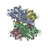
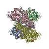
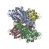
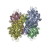
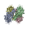
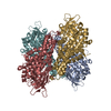
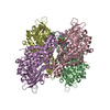
| Title | Crystal structure of FMN-bound DszC from Rhodococcus erythropolis D-1 |
|---|
 Components Components | DszC |
|---|
 Keywords Keywords | OXIDOREDUCTASE / DBT monooxygenase / desulfurization / acyl-CoA dehydrogenase domain / FMN-dependent |
|---|
| Function / homology |  Function and homology information Function and homology information
dibenzothiophene catabolic process / dibenzothiophene monooxygenase / 3-methylbutanoyl-CoA dehydrogenase activity / L-leucine catabolic process / monooxygenase activity / flavin adenine dinucleotide binding / cytoplasmSimilarity search - Function : / Acyl-CoA dehydrogenase, C-terminal domain / Acyl-CoA dehydrogenase, C-terminal domain / Butyryl-Coa Dehydrogenase, subunit A; domain 1 / Acyl-CoA dehydrogenase/oxidase, N-terminal domain / Butyryl-CoA Dehydrogenase, subunit A, domain 2 / Butyryl-CoA Dehydrogenase, subunit A; domain 2 / Acyl-CoA dehydrogenase/oxidase, N-terminal / Acyl-CoA dehydrogenase, N-terminal domain / Acyl-CoA oxidase/dehydrogenase, middle domain ...: / Acyl-CoA dehydrogenase, C-terminal domain / Acyl-CoA dehydrogenase, C-terminal domain / Butyryl-Coa Dehydrogenase, subunit A; domain 1 / Acyl-CoA dehydrogenase/oxidase, N-terminal domain / Butyryl-CoA Dehydrogenase, subunit A, domain 2 / Butyryl-CoA Dehydrogenase, subunit A; domain 2 / Acyl-CoA dehydrogenase/oxidase, N-terminal / Acyl-CoA dehydrogenase, N-terminal domain / Acyl-CoA oxidase/dehydrogenase, middle domain / Acyl-CoA dehydrogenase, middle domain / Acyl-CoA dehydrogenase/oxidase, N-terminal domain superfamily / Butyryl-CoA Dehydrogenase, subunit A, domain 3 / Acyl-CoA oxidase/dehydrogenase, middle domain superfamily / Acyl-CoA dehydrogenase/oxidase, N-terminal and middle domain superfamily / Acyl-CoA dehydrogenase-like, C-terminal / Butyryl-CoA Dehydrogenase, subunit A; domain 3 / Up-down Bundle / Beta Barrel / Orthogonal Bundle / Mainly Beta / Mainly AlphaSimilarity search - Domain/homology |
|---|
| Biological species |  Rhodococcus erythropolis (bacteria) Rhodococcus erythropolis (bacteria) |
|---|
| Method |  X-RAY DIFFRACTION / SYNCHROTRON / MOLECULAR REPLACEMENT / Resolution: 2.3 Å X-RAY DIFFRACTION / SYNCHROTRON / MOLECULAR REPLACEMENT / Resolution: 2.3 Å |
|---|
 Authors Authors | Guan, L.J. / Lee, W.C. / Wang, S.P. / Ohtsuka, J. / Tanokura, M. |
|---|
 Citation Citation | Journal: Febs J. / Year: 2015
Title: Crystal structures of apo-DszC and FMN-bound DszC from Rhodococcus erythropolis D-1.
Authors: Guan, L.J. / Lee, W.C. / Wang, S. / Ohshiro, T. / Izumi, Y. / Ohtsuka, J. / Tanokura, M. |
|---|
| History | | Deposition | Oct 23, 2014 | Deposition site: PDBJ / Processing site: PDBJ |
|---|
| Revision 1.0 | Feb 25, 2015 | Provider: repository / Type: Initial release |
|---|
| Revision 1.1 | Aug 24, 2022 | Group: Database references / Derived calculations
Category: citation / citation_author ...citation / citation_author / database_2 / struct_site
Item: _citation.journal_volume / _citation.page_first ..._citation.journal_volume / _citation.page_first / _citation.page_last / _citation.title / _citation_author.name / _database_2.pdbx_DOI / _database_2.pdbx_database_accession / _struct_site.pdbx_auth_asym_id / _struct_site.pdbx_auth_comp_id / _struct_site.pdbx_auth_seq_id |
|---|
| Revision 1.2 | May 29, 2024 | Group: Data collection / Category: chem_comp_atom / chem_comp_bond |
|---|
|
|---|
 Movie
Movie Controller

 Yorodumi
 Open data
 Basic information
 Structure visualization
 Downloads & links
 Other downloads

 PDBj
PDBj Assembly

 Mass: 456.344 Da / Num. of mol.: 4 / Source method: obtained synthetically / Formula: C17H21N4O9P
 Mass: 18.015 Da / Num. of mol.: 795 / Source method: isolated from a natural source / Formula: H2O
 Sample preparation
 / Beamline: AR-NW12A / Wavelength: 1 Å
 Processing