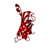
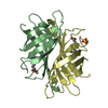
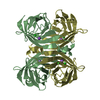
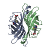
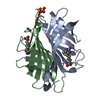
[English] 日本語
Yorodumi
- PDB-3wzn: Crystal structure of the core streptavidin mutant V21 (Y22S/N23D/... -

+
Open data


ID or keywords:

Loading...

-
Basic information

Entry
Database: PDB / ID: 3wzn
TitleCrystal structure of the core streptavidin mutant V21 (Y22S/N23D/S27D/Y83S/R84K/E101D/R103K/E116N) complexed with biotin at 1.3 A resolution
ComponentsStreptavidin
KeywordsBIOTIN BINDING PROTEIN / beta-barrel
Function / homology
Function and homology information


biotin binding / extracellular region
Similarity search - Function
Avidin-like / Avidin-like, conserved site / Avidin-like domain signature. / Avidin / Avidin/streptavidin / Avidin-like superfamily / Avidin family / Avidin-like domain profile. / Lipocalin / Beta Barrel / Mainly Beta
Similarity search - Domain/homology
BIOTIN / Streptavidin
Similarity search - Component
Biological speciesStreptomyces avidinii (bacteria)
MethodX-RAY DIFFRACTION / SYNCHROTRON / MOLECULAR REPLACEMENT / Resolution: 1.3 Å
AuthorsKawato, T. / Mizohata, E. / Shimizu, Y. / Meshizuka, T. / Yamamoto, T. / Takasu, N. / Matsuoka, M. / Matsumura, H. / Tsumoto, K. / Kodama, T. ...Kawato, T. / Mizohata, E. / Shimizu, Y. / Meshizuka, T. / Yamamoto, T. / Takasu, N. / Matsuoka, M. / Matsumura, H. / Tsumoto, K. / Kodama, T. / Kanai, M. / Doi, H. / Inoue, T. / Sugiyama, A.
CitationJournal: J.Biochem. / Year: 2015
Title: Structure-based design of a streptavidin mutant specific for an artificial biotin analogue.
Authors: Kawato, T. / Mizohata, E. / Shimizu, Y. / Meshizuka, T. / Yamamoto, T. / Takasu, N. / Matsuoka, M. / Matsumura, H. / Kodama, T. / Kanai, M. / Doi, H. / Inoue, T. / Sugiyama, A.
History
DepositionOct 1, 2014Deposition site: PDBJ / Processing site: PDBJ
Revision 1.0Feb 18, 2015Provider: repository / Type: Initial release
Revision 1.1Nov 22, 2017Group: Refinement description / Category: software / Item: _software.name
Revision 1.2Aug 24, 2022Group: Database references / Derived calculations
Category: citation / database_2 ...citation / database_2 / struct_ref_seq_dif / struct_site
Item: _citation.journal_volume / _citation.page_first ..._citation.journal_volume / _citation.page_first / _citation.page_last / _citation.title / _database_2.pdbx_DOI / _database_2.pdbx_database_accession / _struct_ref_seq_dif.details / _struct_site.pdbx_auth_asym_id / _struct_site.pdbx_auth_comp_id / _struct_site.pdbx_auth_seq_id
Revision 1.3Nov 8, 2023Group: Data collection / Refinement description
Category: chem_comp_atom / chem_comp_bond / pdbx_initial_refinement_model

-
Structure visualization

Structure viewerMolecule:
MolmilJmol/JSmol

Downloads & links

-
Assembly

Deposited unit
A: Streptavidin
B: Streptavidin
hetero molecules


Theoretical massNumber of molelcules
Total (without water)27,0395
Polymers26,4542
Non-polymers5853
Water3,999222
1
A: Streptavidin
B: Streptavidin
hetero molecules

A: Streptavidin
B: Streptavidin
hetero molecules


Theoretical massNumber of molelcules
Total (without water)54,07810
Polymers52,9094
Non-polymers1,1696
Water724
TypeNameSymmetry operationNumber
identity operation1_555x,y,z1
crystal symmetry operation2_545-x,-y-1,z1
Buried area12020 Å2
ΔGint-63 kcal/mol
Surface area18570 Å2
MethodPISA
Unit cell
Length a, b, c (Å)67.677, 54.344, 60.064
Angle α, β, γ (deg.)90.00, 90.00, 90.00
Int Tables number18
Space group name H-MP21212

-
Components

#1: Protein Streptavidin /


Mass: 13227.240 Da / Num. of mol.: 2 / Fragment: UNP residues 37-163 / Mutation: Y22S/N23D/S27D/Y83S/R84K/E101D/R103K/E116N
Source method: isolated from a genetically manipulated source
Source: (gene. exp.) Streptomyces avidinii (bacteria) / Plasmid: pCold TF / Production host: Escherichia coli (E. coli) / Strain (production host): BL21Star(DE3) / References: UniProt: P22629
#2: Chemical ChemComp-BTN / BIOTIN / Biotin


Mass: 244.311 Da / Num. of mol.: 2 / Source method: obtained synthetically / Formula: C10H16N2O3S
#3: Chemical ChemComp-SO4 / SULFATE ION / Sulfate


Mass: 96.063 Da / Num. of mol.: 1 / Source method: obtained synthetically / Formula: SO4
#4: Water ChemComp-HOH / water / Water


Mass: 18.015 Da / Num. of mol.: 222 / Source method: isolated from a natural source / Formula: H2O

-
Experimental details

-
Experiment

ExperimentMethod: X-RAY DIFFRACTION / Number of used crystals: 1

-
Sample preparation

CrystalDensity Matthews: 2.09 Å3/Da / Density % sol: 41.08 %
Crystal growTemperature: 294 K / Method: vapor diffusion, sitting drop / pH: 4.6
Details: 100 mM sodium acetate trihydrate, 2.0 M ammonium sulfate, pH 4.6, VAPOR DIFFUSION, SITTING DROP, temperature 294K

-
Data collection

DiffractionMean temperature: 100 K
Diffraction sourceSource: SYNCHROTRON / Site: Photon Factory / Beamline: BL-17A / Wavelength: 0.98 Å
DetectorType: ADSC QUANTUM 315r / Detector: CCD / Date: Dec 18, 2012
RadiationProtocol: SINGLE WAVELENGTH / Monochromatic (M) / Laue (L): M / Scattering type: x-ray
Radiation wavelengthWavelength: 0.98 Å / Relative weight: 1
ReflectionResolution: 1.3→50 Å / Num. all: 55326 / Num. obs: 54829 / % possible obs: 99.1 % / Redundancy: 7.5 % / Rsym value: 0.072 / Net I/σ(I): 26.8
Reflection shellResolution: 1.3→1.35 Å / Redundancy: 7.5 % / Mean I/σ(I) obs: 3.95 / Num. unique all: 54829 / Rsym value: 0.406 / % possible all: 99.1

-
Processing

Software
NameVersionClassification
SERGUIdata collection
PHASERphasing
REFMAC5.5.0109refinement
HKL-2000data reduction
HKL-2000data scaling
RefinementMethod to determine structure: MOLECULAR REPLACEMENT
Starting model: 3WYQ
Resolution: 1.3→15 Å / Cor.coef. Fo:Fc: 0.974 / Cor.coef. Fo:Fc free: 0.96 / SU B: 2.288 / SU ML: 0.041 / Cross valid method: THROUGHOUT / ESU R: 0.054 / ESU R Free: 0.055 / Stereochemistry target values: MAXIMUM LIKELIHOOD / Details: HYDROGENS HAVE BEEN ADDED IN THE RIDING POSITIONS
RfactorNum. reflection% reflectionSelection details
Rfree0.20658 2761 5.1 %RANDOM
Rwork0.15995 ---
obs0.16239 51662 98.71 %-
Solvent computationIon probe radii: 0.8 Å / Shrinkage radii: 0.8 Å / VDW probe radii: 1.4 Å / Solvent model: MASK
Displacement parametersBiso mean: 16.37 Å2
Baniso -1Baniso -2Baniso -3
1--0.29 Å20 Å20 Å2
2---0.65 Å20 Å2
3---0.93 Å2
Refinement stepCycle: LAST / Resolution: 1.3→15 Å
ProteinNucleic acidLigandSolventTotal
Num. atoms1801 0 37 222 2060
Refine LS restraints
Refine-IDTypeDev idealDev ideal targetNumber
X-RAY DIFFRACTIONr_bond_refined_d0.0260.0211929
X-RAY DIFFRACTIONr_bond_other_d
X-RAY DIFFRACTIONr_angle_refined_deg2.3281.9162656
X-RAY DIFFRACTIONr_angle_other_deg
X-RAY DIFFRACTIONr_dihedral_angle_1_deg7.1965263
X-RAY DIFFRACTIONr_dihedral_angle_2_deg27.96325.275
X-RAY DIFFRACTIONr_dihedral_angle_3_deg13.6115260
X-RAY DIFFRACTIONr_dihedral_angle_4_deg21.876154
X-RAY DIFFRACTIONr_chiral_restr0.3250.2303
X-RAY DIFFRACTIONr_gen_planes_refined0.0140.021476
X-RAY DIFFRACTIONr_gen_planes_other
X-RAY DIFFRACTIONr_nbd_refined
X-RAY DIFFRACTIONr_nbd_other
X-RAY DIFFRACTIONr_nbtor_refined
X-RAY DIFFRACTIONr_nbtor_other
X-RAY DIFFRACTIONr_xyhbond_nbd_refined
X-RAY DIFFRACTIONr_xyhbond_nbd_other
X-RAY DIFFRACTIONr_metal_ion_refined
X-RAY DIFFRACTIONr_metal_ion_other
X-RAY DIFFRACTIONr_symmetry_vdw_refined
X-RAY DIFFRACTIONr_symmetry_vdw_other
X-RAY DIFFRACTIONr_symmetry_hbond_refined
X-RAY DIFFRACTIONr_symmetry_hbond_other
X-RAY DIFFRACTIONr_symmetry_metal_ion_refined
X-RAY DIFFRACTIONr_symmetry_metal_ion_other
X-RAY DIFFRACTIONr_mcbond_it2.7671.51230
X-RAY DIFFRACTIONr_mcbond_other
X-RAY DIFFRACTIONr_mcangle_it3.82421961
X-RAY DIFFRACTIONr_mcangle_other
X-RAY DIFFRACTIONr_scbond_it5.3413699
X-RAY DIFFRACTIONr_scbond_other
X-RAY DIFFRACTIONr_scangle_it7.1124.5686
X-RAY DIFFRACTIONr_scangle_other
X-RAY DIFFRACTIONr_long_range_B_refined
X-RAY DIFFRACTIONr_long_range_B_other
X-RAY DIFFRACTIONr_rigid_bond_restr2.71931929
X-RAY DIFFRACTIONr_sphericity_free
X-RAY DIFFRACTIONr_sphericity_bonded
LS refinement shellResolution: 1.3→1.334 Å / Total num. of bins used: 20
RfactorNum. reflection% reflection
Rfree0.346 206 -
Rwork0.323 3744 -
obs--98.55 %

+
About Yorodumi

-
News

-
Feb 9, 2022. New format data for meta-information of EMDB entries

New format data for meta-information of EMDB entries

  • Version 3 of the EMDB header file is now the official format.
  • The previous official version 1.9 will be removed from the archive.

Related info.:EMDB header

External links:wwPDB to switch to version 3 of the EMDB data model

-
Aug 12, 2020. Covid-19 info

Covid-19 info

URL: https://pdbj.org/emnavi/covid19.php

New page: Covid-19 featured information page in EM Navigator.

Related info.:Covid-19 info / Mar 5, 2020. Novel coronavirus structure data

+
Mar 5, 2020. Novel coronavirus structure data

Novel coronavirus structure data

Related info.:Yorodumi Speices / Aug 12, 2020. Covid-19 info

External links:COVID-19 featured content - PDBj / Molecule of the Month (242):Coronavirus Proteases

+
Jan 31, 2019. EMDB accession codes are about to change! (news from PDBe EMDB page)

EMDB accession codes are about to change! (news from PDBe EMDB page)

  • The allocation of 4 digits for EMDB accession codes will soon come to an end. Whilst these codes will remain in use, new EMDB accession codes will include an additional digit and will expand incrementally as the available range of codes is exhausted. The current 4-digit format prefixed with “EMD-” (i.e. EMD-XXXX) will advance to a 5-digit format (i.e. EMD-XXXXX), and so on. It is currently estimated that the 4-digit codes will be depleted around Spring 2019, at which point the 5-digit format will come into force.
  • The EM Navigator/Yorodumi systems omit the EMD- prefix.

Related info.:Q: What is EMD? / ID/Accession-code notation in Yorodumi/EM Navigator

External links:EMDB Accession Codes are Changing Soon! / Contact to PDBj

+
Jul 12, 2017. Major update of PDB

Major update of PDB

  • wwPDB released updated PDB data conforming to the new PDBx/mmCIF dictionary.
  • This is a major update changing the version number from 4 to 5, and with Remediation, in which all the entries are updated.
  • In this update, many items about electron microscopy experimental information are reorganized (e.g. em_software).
  • Now, EM Navigator and Yorodumi are based on the updated data.

External links:wwPDB Remediation / Enriched Model Files Conforming to OneDep Data Standards Now Available in the PDB FTP Archive

-
Yorodumi

Thousand views of thousand structures

  • Yorodumi is a browser for structure data from EMDB, PDB, SASBDB, etc.
  • This page is also the successor to EM Navigator detail page, and also detail information page/front-end page for Omokage search.
  • The word "yorodu" (or yorozu) is an old Japanese word meaning "ten thousand". "mi" (miru) is to see.

Related info.:EMDB / PDB / SASBDB / Comparison of 3 databanks / Yorodumi Search / Aug 31, 2016. New EM Navigator & Yorodumi / Yorodumi Papers / Jmol/JSmol / Function and homology information / Changes in new EM Navigator and Yorodumi

Read more