 Movie
Movie Controller

[English] 日本語
 Yorodumi
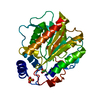
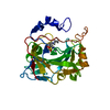
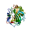
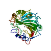
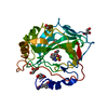
- PDB-3wy6: Structure of hyperthermophilic family 12 endocellulase (E197A) fr... -
+ Open data

- Basic information
| Entry | Database: PDB / ID: 3wy6 | |||||||||
|---|---|---|---|---|---|---|---|---|---|---|
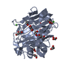
| Title | Structure of hyperthermophilic family 12 endocellulase (E197A) from Pyrococcus furiosus in complex with laminaribiose | |||||||||
 Components Components | Endoglucanase A | |||||||||
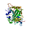
 Keywords Keywords | HYDROLASE / Beta-jelly roll / Glycoside hydrolase | |||||||||
| Function / homology |  Function and homology information Function and homology informationcellulase activity / polysaccharide catabolic process / metal ion binding Similarity search - Function | |||||||||
| Biological species |   Pyrococcus furiosus (archaea) Pyrococcus furiosus (archaea) | |||||||||
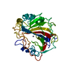
| Method |  X-RAY DIFFRACTION / SYNCHROTRON / MOLECULAR REPLACEMENT / molecular replacement / Resolution: 1.45 Å X-RAY DIFFRACTION / SYNCHROTRON / MOLECULAR REPLACEMENT / molecular replacement / Resolution: 1.45 Å | |||||||||
 Authors Authors | Kataoka, M. / Ishikawa, K. | |||||||||
 Citation Citation | Journal: To be Published Title: Structure of hyperthermophilic family 12 endocellulase (E197A) from Pyrococcus furiosus in complex with laminaribiose Authors: Kataoka, M. / Ishikawa, K. | |||||||||
| History |
|
- Structure visualization
| Structure viewer | Molecule: MolmilJmol/JSmol |
|---|
- Downloads & links
-Download
| PDBx/mmCIF format | 3wy6.cif.gz | 144.2 KB | Display | PDBx/mmCIF format |
|---|---|---|---|---|
| PDB format | pdb3wy6.ent.gz | 111 KB | Display | PDB format |
| PDBx/mmJSON format | 3wy6.json.gz | Tree view | PDBx/mmJSON format | |
| Others |  Other downloads Other downloads |
-Validation report
| Arichive directory | https://data.pdbj.org/pub/pdb/validation_reports/wy/3wy6ftp://data.pdbj.org/pub/pdb/validation_reports/wy/3wy6 | HTTPS FTP |
|---|
-Related structure data
| Related structure data |  3wxd 3wxh 3wxn |
|---|---|
| Similar structure data |


-Links
 PDBj
PDBj


- Assembly
| Deposited unit | 
| ||||||||
|---|---|---|---|---|---|---|---|---|---|
| 1 |
| ||||||||
| Unit cell |
| ||||||||
| Components on special symmetry positions |
|
-Components
-Protein , 1 types, 1 molecules A
| #1: Protein | Mass: 35957.543 Da / Num. of mol.: 1 / Mutation: E197A Source method: isolated from a genetically manipulated source Source: (gene. exp.) Pyrococcus furiosus (archaea) / Gene: eglA / Production host:  |
|---|
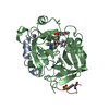
-Sugars , 2 types, 2 molecules 
| #2: Polysaccharide | beta-D-glucopyranose-(1-3)-beta-D-glucopyranose / beta-laminaribiose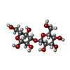  Source method: isolated from a genetically manipulated source Details: oligosaccharide / References: beta-laminaribiose |
|---|---|
| #5: Sugar | ChemComp-BGC /  Type: D-saccharide, beta linking / Mass: 180.156 Da / Num. of mol.: 1 Type: D-saccharide, beta linking / Mass: 180.156 Da / Num. of mol.: 1Source method: isolated from a genetically manipulated source Formula: C6H12O6 |
-Non-polymers , 4 types, 254 molecules 


| #3: Chemical |  Mass: 40.078 Da / Num. of mol.: 2 / Source method: obtained synthetically / Formula: Ca Mass: 40.078 Da / Num. of mol.: 2 / Source method: obtained synthetically / Formula: Ca#4: Chemical | ChemComp-NHE /  Mass: 207.290 Da / Num. of mol.: 4 / Source method: obtained synthetically / Formula: C8H17NO3S / Comment: pH buffer*YM Mass: 207.290 Da / Num. of mol.: 4 / Source method: obtained synthetically / Formula: C8H17NO3S / Comment: pH buffer*YM#6: Chemical | ChemComp-GOL /  Mass: 92.094 Da / Num. of mol.: 8 / Source method: obtained synthetically / Formula: C3H8O3 Mass: 92.094 Da / Num. of mol.: 8 / Source method: obtained synthetically / Formula: C3H8O3#7: Water | ChemComp-HOH / | Mass: 18.015 Da / Num. of mol.: 240 / Source method: isolated from a natural source / Formula: H2O |
|---|
-Experimental details
-Experiment
| Experiment | Method: X-RAY DIFFRACTION / Number of used crystals: 1 |
|---|
- Sample preparation
| Crystal | Density Matthews: 2.25 Å3/Da / Density % sol: 45.28 % / Mosaicity: 0.447 ° / Mosaicity esd: 0.004 ° |
|---|---|
| Crystal grow | Temperature: 303 K / Method: vapor diffusion, hanging drop / pH: 9 Details: 0.12M CHES, 0.5M potassium sodium tartrate, 0.1M lithium sulfate, pH 9.0, VAPOR DIFFUSION, HANGING DROP, temperature 303.0K |
-Data collection
| Diffraction | Mean temperature: 100 K | ||||||||||||||||||||||||||||||||||||||||||||||||||||||||||||||||||||||||||||||||||||||||||||||||||||||||||||||||||||||||||||||
|---|---|---|---|---|---|---|---|---|---|---|---|---|---|---|---|---|---|---|---|---|---|---|---|---|---|---|---|---|---|---|---|---|---|---|---|---|---|---|---|---|---|---|---|---|---|---|---|---|---|---|---|---|---|---|---|---|---|---|---|---|---|---|---|---|---|---|---|---|---|---|---|---|---|---|---|---|---|---|---|---|---|---|---|---|---|---|---|---|---|---|---|---|---|---|---|---|---|---|---|---|---|---|---|---|---|---|---|---|---|---|---|---|---|---|---|---|---|---|---|---|---|---|---|---|---|---|---|
| Diffraction source | Source: SYNCHROTRON / Site: SPring-8  / Beamline: BL44XU / Wavelength: 0.9 Å / Beamline: BL44XU / Wavelength: 0.9 Å | ||||||||||||||||||||||||||||||||||||||||||||||||||||||||||||||||||||||||||||||||||||||||||||||||||||||||||||||||||||||||||||||
| Detector | Type: RAYONIX MX225HE / Detector: CCD / Date: Oct 17, 2012 | ||||||||||||||||||||||||||||||||||||||||||||||||||||||||||||||||||||||||||||||||||||||||||||||||||||||||||||||||||||||||||||||
| Radiation | Monochromator: double-crystal / Protocol: SINGLE WAVELENGTH / Monochromatic (M) / Laue (L): M / Scattering type: x-ray | ||||||||||||||||||||||||||||||||||||||||||||||||||||||||||||||||||||||||||||||||||||||||||||||||||||||||||||||||||||||||||||||
| Radiation wavelength | Wavelength: 0.9 Å / Relative weight: 1 | ||||||||||||||||||||||||||||||||||||||||||||||||||||||||||||||||||||||||||||||||||||||||||||||||||||||||||||||||||||||||||||||
| Reflection | Resolution: 1.45→50 Å / Num. obs: 55555 / % possible obs: 95.6 % / Redundancy: 3.7 % / Rmerge(I) obs: 0.07 / Χ2: 0.981 / Net I/σ(I): 11.2 | ||||||||||||||||||||||||||||||||||||||||||||||||||||||||||||||||||||||||||||||||||||||||||||||||||||||||||||||||||||||||||||||
| Reflection shell | Diffraction-ID: 1 / Rejects: _
|
-Phasing
| Phasing | Method: molecular replacement |
|---|
- Processing
| Software |
| ||||||||||||||||||||||||||||||||||||||||||||||||||||||||||||
|---|---|---|---|---|---|---|---|---|---|---|---|---|---|---|---|---|---|---|---|---|---|---|---|---|---|---|---|---|---|---|---|---|---|---|---|---|---|---|---|---|---|---|---|---|---|---|---|---|---|---|---|---|---|---|---|---|---|---|---|---|---|
| Refinement | Method to determine structure: MOLECULAR REPLACEMENT / Resolution: 1.45→41.55 Å / Cor.coef. Fo:Fc: 0.978 / Cor.coef. Fo:Fc free: 0.963 / WRfactor Rfree: 0.1669 / WRfactor Rwork: 0.1208 / FOM work R set: 0.907 / SU B: 2.387 / SU ML: 0.039 / SU R Cruickshank DPI: 0.0564 / SU Rfree: 0.0595 / Cross valid method: THROUGHOUT / σ(F): 0 / ESU R: 0.056 / ESU R Free: 0.059 / Stereochemistry target values: MAXIMUM LIKELIHOOD Details: HYDROGENS HAVE BEEN USED IF PRESENT IN THE INPUT U VALUES
| ||||||||||||||||||||||||||||||||||||||||||||||||||||||||||||
| Solvent computation | Ion probe radii: 0.8 Å / Shrinkage radii: 0.8 Å / VDW probe radii: 1.2 Å / Solvent model: MASK | ||||||||||||||||||||||||||||||||||||||||||||||||||||||||||||
| Displacement parameters | Biso max: 124.47 Å2 / Biso mean: 16.696 Å2 / Biso min: 5.27 Å2
| ||||||||||||||||||||||||||||||||||||||||||||||||||||||||||||
| Refinement step | Cycle: LAST / Resolution: 1.45→41.55 Å
| ||||||||||||||||||||||||||||||||||||||||||||||||||||||||||||
| Refine LS restraints |
| ||||||||||||||||||||||||||||||||||||||||||||||||||||||||||||
| LS refinement shell | Resolution: 1.451→1.488 Å / Total num. of bins used: 20
|
