| Entry | Database: PDB / ID: 3wr7
|
|---|
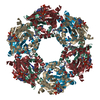
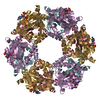
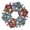
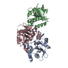
| Title | Crystal Structure of Spermidine Acetyltransferase from Escherichia coli |
|---|
 Components Components | Spermidine N1-acetyltransferase |
|---|
 Keywords Keywords | TRANSFERASE / alpha and beta |
|---|
| Function / homology |  Function and homology information Function and homology information
Acetyltransferase (GNAT) domain / Gcn5-related N-acetyltransferase (GNAT) / Gcn5-related N-acetyltransferase (GNAT) domain profile. / GNAT domain / Acyl-CoA N-acyltransferase / Aminopeptidase / 3-Layer(aba) Sandwich / Alpha BetaSimilarity search - Domain/homology |
|---|
| Biological species |   Escherichia coli (E. coli) Escherichia coli (E. coli) |
|---|
| Method |  X-RAY DIFFRACTION / SYNCHROTRON / MOLECULAR REPLACEMENT / Resolution: 2.5 Å X-RAY DIFFRACTION / SYNCHROTRON / MOLECULAR REPLACEMENT / Resolution: 2.5 Å |
|---|
 Authors Authors | Sugiyama, S. / Ishikawa, S. / Tomitori, S. / Niiyama, M. / Hirose, M. / Miyazaki, Y. / Higashi, K. / Adachi, H. / Takano, K. / Murakami, S. ...Sugiyama, S. / Ishikawa, S. / Tomitori, S. / Niiyama, M. / Hirose, M. / Miyazaki, Y. / Higashi, K. / Adachi, H. / Takano, K. / Murakami, S. / Inoue, T. / Mori, Y. / Kashiwagi, K. / Igarashi, K. / Matsumura, H. |
|---|
 Citation Citation | Journal: Int.J.Biochem.Cell Biol. / Year: 2016
Title: Molecular mechanism underlying promiscuous polyamine recognition by spermidine acetyltransferase
Authors: Sugiyama, S. / Ishikawa, S. / Tomitori, H. / Niiyama, M. / Hirose, M. / Miyazaki, Y. / Higashi, K. / Murata, M. / Adachi, H. / Takano, K. / Murakami, S. / Inoue, T. / Mori, Y. / Kashiwagi, K. ...Authors: Sugiyama, S. / Ishikawa, S. / Tomitori, H. / Niiyama, M. / Hirose, M. / Miyazaki, Y. / Higashi, K. / Murata, M. / Adachi, H. / Takano, K. / Murakami, S. / Inoue, T. / Mori, Y. / Kashiwagi, K. / Igarashi, K. / Matsumura, H. |
|---|
| History | | Deposition | Feb 20, 2014 | Deposition site: PDBJ / Processing site: PDBJ |
|---|
| Revision 1.0 | Sep 2, 2015 | Provider: repository / Type: Initial release |
|---|
| Revision 1.1 | Jun 8, 2016 | Group: Database references |
|---|
| Revision 1.2 | Mar 20, 2024 | Group: Data collection / Database references / Derived calculations
Category: chem_comp_atom / chem_comp_bond ...chem_comp_atom / chem_comp_bond / database_2 / struct_site
Item: _database_2.pdbx_DOI / _database_2.pdbx_database_accession ..._database_2.pdbx_DOI / _database_2.pdbx_database_accession / _struct_site.pdbx_auth_asym_id / _struct_site.pdbx_auth_comp_id / _struct_site.pdbx_auth_seq_id |
|---|
|
|---|
 Movie
Movie Controller

 Yorodumi
 Open data
 Basic information
 Structure visualization
 Downloads & links
 Other downloads


 PDBj
PDBj


 Assembly

 Mass: 145.246 Da / Num. of mol.: 4 / Source method: obtained synthetically / Formula: C7H19N3
 Mass: 767.534 Da / Num. of mol.: 4 / Source method: obtained synthetically / Formula: C21H36N7O16P3S
 Mass: 18.015 Da / Num. of mol.: 170 / Source method: isolated from a natural source / Formula: H2O
 Sample preparation
 / Beamline: BL41XU / Wavelength: 0.97 Å
 Processing