- PDB-3wnz: Crystal structure of Bacillus subtilis YwfE, an L-amino acid liga... -
+
Open data
ID or keywords:
Loading...
-
Basic information
Entry
Database: PDB / ID: 3wnz
Title
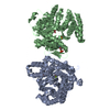
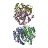
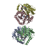
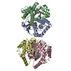
Crystal structure of Bacillus subtilis YwfE, an L-amino acid ligase, with bound ADP-Mg-Pi
Components
Alanine-anticapsin ligase BacD
Keywords
LIGASE / ATP-grasp Fold / ATP binding
Function / homology
Function and homology information
L-alanine-L-anticapsin ligase / L-amino-acid alpha-ligase activity / antibiotic biosynthetic process / ATP binding / metal ion binding Similarity search - Function
Monochromator: Si 111 / Protocol: SINGLE WAVELENGTH / Monochromatic (M) / Laue (L): M / Scattering type: x-ray
Radiation wavelength
Wavelength: 1 Å / Relative weight: 1
Reflection
Resolution: 1.9→20 Å / Num. obs: 43922 / % possible obs: 88.9 % / Redundancy: 10.6 % / Rmerge(I) obs: 0.069 / Net I/σ(I): 53.5
Reflection shell
Resolution: 1.9→1.95 Å / Redundancy: 11.1 % / Rmerge(I) obs: 0.312 / Mean I/σ(I) obs: 9.9 / Num. unique all: 3319 / % possible all: 96.5
-
Processing
Software
Name
Version
Classification
HKL-2000
datacollection
CRANK
phasing
REFMAC
5.7.0029
refinement
HKL-2000
datareduction
HKL-2000
datascaling
Refinement
Method to determine structure: SAD / Resolution: 1.9→17.42 Å / Cor.coef. Fo:Fc: 0.944 / Cor.coef. Fo:Fc free: 0.914 / SU B: 7.238 / SU ML: 0.116 / Cross valid method: THROUGHOUT / ESU R: 0.158 / ESU R Free: 0.153 / Stereochemistry target values: MAXIMUM LIKELIHOOD / Details: HYDROGENS HAVE BEEN ADDED IN THE RIDING POSITIONS
Rfactor
Num. reflection
% reflection
Selection details
Rfree
0.24301
2232
5.1 %
RANDOM
Rwork
0.19658
-
-
-
obs
0.19899
41606
88.87 %
-
Solvent computation
Ion probe radii: 0.8 Å / Shrinkage radii: 0.8 Å / VDW probe radii: 1.2 Å / Solvent model: MASK
 Movie
Movie Controller

 Yorodumi
 Open data
 Basic information
 Components
 Keywords
 Function and homology information
 X-RAY DIFFRACTION /
  Authors
 Citation
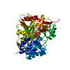
 Structure visualization
 Downloads & links
 Other downloads
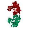


 PDBj
PDBj


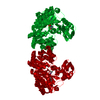
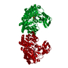
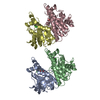
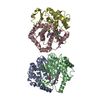
 Assembly
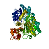


 Mass: 427.201 Da / Num. of mol.: 1 / Source method: obtained synthetically / Formula: C10H15N5O10P2 / Comment: ADP, energy-carrying molecule*YM
 Mass: 24.305 Da / Num. of mol.: 2 / Source method: obtained synthetically / Formula: Mg
 Mass: 94.971 Da / Num. of mol.: 1 / Source method: obtained synthetically / Formula: PO4
 Mass: 18.015 Da / Num. of mol.: 396 / Source method: isolated from a natural source / Formula: H2O
 Sample preparation
 / Beamline: BL-5A / Wavelength: 1 Å
 Processing