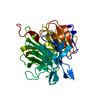
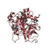
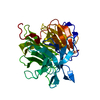
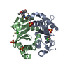
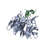
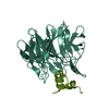
[English] 日本語
Yorodumi
- PDB-3wn7: Crystal Structure of Keap1 in Complex with the N-terminal region ... -

+
Open data


ID or keywords:

Loading...

-
Basic information

Entry
Database: PDB / ID: 3wn7
TitleCrystal Structure of Keap1 in Complex with the N-terminal region of the Nrf2 transcription factor
Components
  • Kelch-like ECH-associated protein 1
  • Peptide from Nuclear factor erythroid 2-related factor 2
KeywordsTRANSCRIPTION / beta-propeller / Kelch motif / Degron
Function / homology
Function and homology information


positive regulation of glutathione biosynthetic process / aflatoxin catabolic process / negative regulation of hematopoietic stem cell differentiation / Nuclear events mediated by NFE2L2 / GSK3B and BTRC:CUL1-mediated-degradation of NFE2L2 / negative regulation of cellular response to hypoxia / PERK-mediated unfolded protein response / cellular response to carbohydrate stimulus / regulation of cellular response to oxidative stress / regulation of removal of superoxide radicals ...positive regulation of glutathione biosynthetic process / aflatoxin catabolic process / negative regulation of hematopoietic stem cell differentiation / Nuclear events mediated by NFE2L2 / GSK3B and BTRC:CUL1-mediated-degradation of NFE2L2 / negative regulation of cellular response to hypoxia / PERK-mediated unfolded protein response / cellular response to carbohydrate stimulus / regulation of cellular response to oxidative stress / regulation of removal of superoxide radicals / KEAP1-NFE2L2 pathway / negative regulation of vascular associated smooth muscle cell migration / regulation of epidermal cell differentiation / Neddylation / cellular response to laminar fluid shear stress / Antigen processing: Ubiquitination & Proteasome degradation / Ub-specific processing proteases / cellular response to fluid shear stress / cellular response to angiotensin / negative regulation of cardiac muscle cell apoptotic process / regulation of innate immune response / Cul3-RING ubiquitin ligase complex / proteasomal ubiquitin-independent protein catabolic process / regulation of embryonic development / transcription factor binding / ubiquitin-like ligase-substrate adaptor activity / centriolar satellite / positive regulation of blood vessel endothelial cell migration / cellular response to glucose starvation / positive regulation of blood coagulation / negative regulation of endothelial cell apoptotic process / negative regulation of oxidative stress-induced intrinsic apoptotic signaling pathway / inclusion body / endoplasmic reticulum unfolded protein response / cellular response to copper ion / cellular response to interleukin-4 / response to endoplasmic reticulum stress / cell redox homeostasis / response to organic substance / regulation of autophagy / actin filament / response to ischemia / transcription coregulator binding / protein-DNA complex / adherens junction / positive regulation of glucose import / positive regulation of neuron projection development / cellular response to hydrogen peroxide / RNA polymerase II transcription regulator complex / positive regulation of angiogenesis / positive regulation of reactive oxygen species metabolic process / disordered domain specific binding / cellular response to xenobiotic stimulus / cellular response to oxidative stress / cellular response to tumor necrosis factor / midbody / ubiquitin-dependent protein catabolic process / cellular response to hypoxia / proteasome-mediated ubiquitin-dependent protein catabolic process / DNA-binding transcription activator activity, RNA polymerase II-specific / response to oxidative stress / in utero embryonic development / transcription regulator complex / RNA polymerase II-specific DNA-binding transcription factor binding / sequence-specific DNA binding / protein ubiquitination / transcription cis-regulatory region binding / DNA-binding transcription factor activity, RNA polymerase II-specific / inflammatory response / RNA polymerase II cis-regulatory region sequence-specific DNA binding / DNA-binding transcription factor activity / protein domain specific binding / negative regulation of gene expression / focal adhesion / centrosome / ubiquitin protein ligase binding / chromatin / positive regulation of gene expression / regulation of DNA-templated transcription / regulation of transcription by RNA polymerase II / positive regulation of DNA-templated transcription / Golgi apparatus / endoplasmic reticulum / positive regulation of transcription by RNA polymerase II / protein-containing complex / DNA binding / nucleoplasm / identical protein binding / nucleus / plasma membrane / cytosol / cytoplasm
Similarity search - Function
: / Basic leucine zipper domain, Maf-type / bZIP Maf transcription factor / Transcription factor, Skn-1-like, DNA-binding domain superfamily / Kelch-type beta propeller / Kelch-like ECH-associated protein 1 / : / BTB-kelch protein / BTB/Kelch-associated / BTB And C-terminal Kelch ...: / Basic leucine zipper domain, Maf-type / bZIP Maf transcription factor / Transcription factor, Skn-1-like, DNA-binding domain superfamily / Kelch-type beta propeller / Kelch-like ECH-associated protein 1 / : / BTB-kelch protein / BTB/Kelch-associated / BTB And C-terminal Kelch / BTB And C-terminal Kelch / Kelch / Kelch repeat type 1 / Kelch motif / Basic-leucine zipper (bZIP) domain signature. / Basic-leucine zipper (bZIP) domain profile. / basic region leucin zipper / Basic-leucine zipper domain superfamily / Basic-leucine zipper domain / Kelch-type beta propeller / BTB/POZ domain / BTB domain profile. / Broad-Complex, Tramtrack and Bric a brac / BTB/POZ domain / 6 Propeller / Neuraminidase / SKP1/BTB/POZ domain superfamily / Mainly Beta
Similarity search - Domain/homology
ACETATE ION / Nuclear factor erythroid 2-related factor 2 / Kelch-like ECH-associated protein 1
Similarity search - Component
Biological speciesMus musculus (house mouse)
MethodX-RAY DIFFRACTION / SYNCHROTRON / MOLECULAR REPLACEMENT / Resolution: 1.57 Å
AuthorsFukutomi, T. / Takagi, K. / Mizushima, T. / Ohuchi, N. / Yamamoto, M.
CitationJournal: Mol.Cell.Biol. / Year: 2014
Title: Kinetic, thermodynamic, and structural characterizations of the association between Nrf2-DLGex degron and Keap1
Authors: Fukutomi, T. / Takagi, K. / Mizushima, T. / Ohuchi, N. / Yamamoto, M.
History
DepositionDec 5, 2013Deposition site: PDBJ / Processing site: PDBJ
Revision 1.0Dec 25, 2013Provider: repository / Type: Initial release
Revision 1.1Jul 16, 2014Group: Database references
Revision 1.2Nov 8, 2023Group: Data collection / Database references ...Data collection / Database references / Derived calculations / Refinement description
Category: chem_comp_atom / chem_comp_bond ...chem_comp_atom / chem_comp_bond / database_2 / pdbx_initial_refinement_model / struct_ref_seq_dif / struct_site
Item: _database_2.pdbx_DOI / _database_2.pdbx_database_accession ..._database_2.pdbx_DOI / _database_2.pdbx_database_accession / _struct_ref_seq_dif.details / _struct_site.pdbx_auth_asym_id / _struct_site.pdbx_auth_comp_id / _struct_site.pdbx_auth_seq_id

-
Structure visualization

Structure viewerMolecule:
MolmilJmol/JSmol

Downloads & links

-
Assembly

Deposited unit
A: Kelch-like ECH-associated protein 1
B: Peptide from Nuclear factor erythroid 2-related factor 2
L: Kelch-like ECH-associated protein 1
M: Peptide from Nuclear factor erythroid 2-related factor 2
hetero molecules


Theoretical massNumber of molelcules
Total (without water)77,3286
Polymers77,2104
Non-polymers1182
Water8,809489
1
A: Kelch-like ECH-associated protein 1
B: Peptide from Nuclear factor erythroid 2-related factor 2
hetero molecules


Theoretical massNumber of molelcules
Total (without water)38,6643
Polymers38,6052
Non-polymers591
Water362
TypeNameSymmetry operationNumber
identity operation1_555x,y,z1
Buried area1680 Å2
ΔGint-6 kcal/mol
Surface area12730 Å2
MethodPISA
2
L: Kelch-like ECH-associated protein 1
M: Peptide from Nuclear factor erythroid 2-related factor 2
hetero molecules


Theoretical massNumber of molelcules
Total (without water)38,6643
Polymers38,6052
Non-polymers591
Water362
TypeNameSymmetry operationNumber
identity operation1_555x,y,z1
Buried area1690 Å2
ΔGint-5 kcal/mol
Surface area12600 Å2
MethodPISA
Unit cell
Length a, b, c (Å)66.138, 84.394, 69.818
Angle α, β, γ (deg.)90.00, 116.89, 90.00
Int Tables number4
Space group name H-MP1211

-
Components

#1: Protein Kelch-like ECH-associated protein 1 / Cytosolic inhibitor of Nrf2 / INrf2


Mass: 34270.109 Da / Num. of mol.: 2 / Fragment: Keap1-DC, UNP residues 321-609
Source method: isolated from a genetically manipulated source
Source: (gene. exp.) Mus musculus (house mouse) / Gene: Keap1, Inrf2, Kiaa0132 / Plasmid: pET101 / Production host: Escherichia coli (E. coli) / Strain (production host): BL 21 Gold(DE3) / References: UniProt: Q9Z2X8
#2: Protein/peptide Peptide from Nuclear factor erythroid 2-related factor 2 / / NF-E2-related factor 2 / NFE2-related factor 2 / Nuclear factor / erythroid derived 2 / like 2


Mass: 4334.840 Da / Num. of mol.: 2 / Fragment: UNP residues 17-51 / Source method: obtained synthetically / Source: (synth.) Mus musculus (house mouse) / References: UniProt: Q60795
#3: Chemical ChemComp-ACT / ACETATE ION / Acetate


Mass: 59.044 Da / Num. of mol.: 2 / Source method: obtained synthetically / Formula: C2H3O2
#4: Water ChemComp-HOH / water / Water


Mass: 18.015 Da / Num. of mol.: 489 / Source method: isolated from a natural source / Formula: H2O

-
Experimental details

-
Experiment

ExperimentMethod: X-RAY DIFFRACTION / Number of used crystals: 1

-
Sample preparation

CrystalDensity Matthews: 2.25 Å3/Da / Density % sol: 45.35 %
Crystal growTemperature: 277 K / Method: vapor diffusion, sitting drop / pH: 7.6
Details: 100mM Sodium HEPES, 0.2M potassium acetate, 20% (w/v) PEG 3350, pH 7.6, VAPOR DIFFUSION, SITTING DROP, temperature 277.0K

-
Data collection

DiffractionMean temperature: 100 K
Diffraction sourceSource: SYNCHROTRON / Site: SPring-8 / Beamline: BL44XU / Wavelength: 0.9 Å
DetectorType: RAYONIX MX300HE / Detector: CCD / Date: Nov 24, 2013
RadiationProtocol: SINGLE WAVELENGTH / Monochromatic (M) / Laue (L): M / Scattering type: x-ray
Radiation wavelengthWavelength: 0.9 Å / Relative weight: 1
ReflectionResolution: 1.57→50 Å / Num. obs: 93450 / % possible obs: 98.8 % / Observed criterion σ(I): -3 / Redundancy: 3.7 % / Rmerge(I) obs: 0.097 / Net I/σ(I): 18.6
Reflection shellResolution: 1.57→1.61 Å / Redundancy: 3.4 % / Rmerge(I) obs: 0.253 / Mean I/σ(I) obs: 7.8 / % possible all: 88.7

-
Processing

Software
NameVersionClassification
HKL-2000data collection
MOLREPphasing
REFMAC5.6.0117refinement
HKL-2000data reduction
HKL-2000data scaling
RefinementMethod to determine structure: MOLECULAR REPLACEMENT
Starting model: PDB ENTRY 1X2J
Resolution: 1.57→34.78 Å / Cor.coef. Fo:Fc: 0.955 / Cor.coef. Fo:Fc free: 0.938 / SU B: 1.421 / SU ML: 0.052 / Cross valid method: THROUGHOUT / ESU R: 0.087 / ESU R Free: 0.089 / Stereochemistry target values: MAXIMUM LIKELIHOOD / Details: HYDROGENS HAVE BEEN USED IF PRESENT IN THE INPUT
RfactorNum. reflection% reflectionSelection details
Rfree0.22299 4688 5 %RANDOM
Rwork0.18749 ---
obs0.18923 88724 98.64 %-
Solvent computationIon probe radii: 0.8 Å / Shrinkage radii: 0.8 Å / VDW probe radii: 1.2 Å / Solvent model: MASK
Displacement parametersBiso mean: 17.864 Å2
Baniso -1Baniso -2Baniso -3
1-0.01 Å2-0 Å20 Å2
2---0.01 Å2-0 Å2
3---0 Å2
Refinement stepCycle: LAST / Resolution: 1.57→34.78 Å
ProteinNucleic acidLigandSolventTotal
Num. atoms4796 0 8 489 5293
Refine LS restraints
Refine-IDTypeDev idealDev ideal targetNumber
X-RAY DIFFRACTIONr_bond_refined_d0.0250.0195176
X-RAY DIFFRACTIONr_bond_other_d
X-RAY DIFFRACTIONr_angle_refined_deg2.3431.9367090
X-RAY DIFFRACTIONr_angle_other_deg
X-RAY DIFFRACTIONr_dihedral_angle_1_deg7.2615683
X-RAY DIFFRACTIONr_dihedral_angle_2_deg34.00322.713247
X-RAY DIFFRACTIONr_dihedral_angle_3_deg13.21415796
X-RAY DIFFRACTIONr_dihedral_angle_4_deg17.3311550
X-RAY DIFFRACTIONr_chiral_restr0.1930.2762
X-RAY DIFFRACTIONr_gen_planes_refined0.0150.0214118
X-RAY DIFFRACTIONr_gen_planes_other
X-RAY DIFFRACTIONr_nbd_refined
X-RAY DIFFRACTIONr_nbd_other
X-RAY DIFFRACTIONr_nbtor_refined
X-RAY DIFFRACTIONr_nbtor_other
X-RAY DIFFRACTIONr_xyhbond_nbd_refined
X-RAY DIFFRACTIONr_xyhbond_nbd_other
X-RAY DIFFRACTIONr_metal_ion_refined
X-RAY DIFFRACTIONr_metal_ion_other
X-RAY DIFFRACTIONr_symmetry_vdw_refined
X-RAY DIFFRACTIONr_symmetry_vdw_other
X-RAY DIFFRACTIONr_symmetry_hbond_refined
X-RAY DIFFRACTIONr_symmetry_hbond_other
X-RAY DIFFRACTIONr_symmetry_metal_ion_refined
X-RAY DIFFRACTIONr_symmetry_metal_ion_other
X-RAY DIFFRACTIONr_mcbond_it
X-RAY DIFFRACTIONr_mcbond_other
X-RAY DIFFRACTIONr_mcangle_it
X-RAY DIFFRACTIONr_mcangle_other
X-RAY DIFFRACTIONr_scbond_it
X-RAY DIFFRACTIONr_scbond_other
X-RAY DIFFRACTIONr_scangle_it
X-RAY DIFFRACTIONr_scangle_other
X-RAY DIFFRACTIONr_long_range_B_refined
X-RAY DIFFRACTIONr_long_range_B_other
X-RAY DIFFRACTIONr_rigid_bond_restr
X-RAY DIFFRACTIONr_sphericity_free
X-RAY DIFFRACTIONr_sphericity_bonded
LS refinement shellResolution: 1.574→1.614 Å / Total num. of bins used: 20
RfactorNum. reflection% reflection
Rfree0.262 336 -
Rwork0.213 5752 -
obs--88.54 %

+
About Yorodumi

-
News

-
Feb 9, 2022. New format data for meta-information of EMDB entries

New format data for meta-information of EMDB entries

  • Version 3 of the EMDB header file is now the official format.
  • The previous official version 1.9 will be removed from the archive.

Related info.:EMDB header

External links:wwPDB to switch to version 3 of the EMDB data model

-
Aug 12, 2020. Covid-19 info

Covid-19 info

URL: https://pdbj.org/emnavi/covid19.php

New page: Covid-19 featured information page in EM Navigator.

Related info.:Covid-19 info / Mar 5, 2020. Novel coronavirus structure data

+
Mar 5, 2020. Novel coronavirus structure data

Novel coronavirus structure data

Related info.:Yorodumi Speices / Aug 12, 2020. Covid-19 info

External links:COVID-19 featured content - PDBj / Molecule of the Month (242):Coronavirus Proteases

+
Jan 31, 2019. EMDB accession codes are about to change! (news from PDBe EMDB page)

EMDB accession codes are about to change! (news from PDBe EMDB page)

  • The allocation of 4 digits for EMDB accession codes will soon come to an end. Whilst these codes will remain in use, new EMDB accession codes will include an additional digit and will expand incrementally as the available range of codes is exhausted. The current 4-digit format prefixed with “EMD-” (i.e. EMD-XXXX) will advance to a 5-digit format (i.e. EMD-XXXXX), and so on. It is currently estimated that the 4-digit codes will be depleted around Spring 2019, at which point the 5-digit format will come into force.
  • The EM Navigator/Yorodumi systems omit the EMD- prefix.

Related info.:Q: What is EMD? / ID/Accession-code notation in Yorodumi/EM Navigator

External links:EMDB Accession Codes are Changing Soon! / Contact to PDBj

+
Jul 12, 2017. Major update of PDB

Major update of PDB

  • wwPDB released updated PDB data conforming to the new PDBx/mmCIF dictionary.
  • This is a major update changing the version number from 4 to 5, and with Remediation, in which all the entries are updated.
  • In this update, many items about electron microscopy experimental information are reorganized (e.g. em_software).
  • Now, EM Navigator and Yorodumi are based on the updated data.

External links:wwPDB Remediation / Enriched Model Files Conforming to OneDep Data Standards Now Available in the PDB FTP Archive

-
Yorodumi

Thousand views of thousand structures

  • Yorodumi is a browser for structure data from EMDB, PDB, SASBDB, etc.
  • This page is also the successor to EM Navigator detail page, and also detail information page/front-end page for Omokage search.
  • The word "yorodu" (or yorozu) is an old Japanese word meaning "ten thousand". "mi" (miru) is to see.

Related info.:EMDB / PDB / SASBDB / Comparison of 3 databanks / Yorodumi Search / Aug 31, 2016. New EM Navigator & Yorodumi / Yorodumi Papers / Jmol/JSmol / Function and homology information / Changes in new EM Navigator and Yorodumi

Read more