 Movie
Movie Controller

[English] 日本語
 Yorodumi
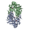
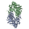
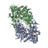
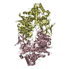
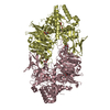
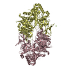
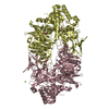
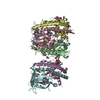
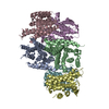
- PDB-3wfz: Crystal structure of Galacto-N-Biose/Lacto-N-Biose I Phosphorylas... -
+ Open data

- Basic information
| Entry | Database: PDB / ID: 3wfz | |||||||||
|---|---|---|---|---|---|---|---|---|---|---|
| Title | Crystal structure of Galacto-N-Biose/Lacto-N-Biose I Phosphorylase C236Y Mutant | |||||||||
 Components Components | Lacto-N-biose phosphorylase | |||||||||
 Keywords Keywords | TRANSFERASE / beta-alpha-barrel / TIM barrel / phosphorylase | |||||||||
| Function / homology |  Function and homology information Function and homology information1,3-beta-galactosyl-N-acetylhexosamine phosphorylase / 1,3-beta-galactosyl-N-acetylhexosamine phosphorylase activity / 1,4-alpha-oligoglucan phosphorylase activity / carbohydrate metabolic process Similarity search - Function | |||||||||
| Biological species |  Bifidobacterium longum (bacteria) Bifidobacterium longum (bacteria) | |||||||||
| Method |  X-RAY DIFFRACTION / SYNCHROTRON / MOLECULAR REPLACEMENT / Resolution: 2.6 Å X-RAY DIFFRACTION / SYNCHROTRON / MOLECULAR REPLACEMENT / Resolution: 2.6 Å | |||||||||
 Authors Authors | Koyama, Y. / Hidaka, M. / Kawakami, M. / Nishimoto, M. / Kitaoka, M. | |||||||||
 Citation Citation | Journal: Protein Eng.Des.Sel. / Year: 2013 Title: Directed evolution to enhance thermostability of galacto-N-biose/lacto-N-biose I phosphorylase. Authors: Koyama, Y. / Hidaka, M. / Nishimoto, M. / Kitaoka, M. | |||||||||
| History |
|
- Structure visualization
| Structure viewer | Molecule: MolmilJmol/JSmol |
|---|
- Downloads & links
-Download
| PDBx/mmCIF format | 3wfz.cif.gz | 591.6 KB | Display | PDBx/mmCIF format |
|---|---|---|---|---|
| PDB format | pdb3wfz.ent.gz | 485.7 KB | Display | PDB format |
| PDBx/mmJSON format | 3wfz.json.gz | Tree view | PDBx/mmJSON format | |
| Others |  Other downloads Other downloads |
-Validation report
| Arichive directory | https://data.pdbj.org/pub/pdb/validation_reports/wf/3wfzftp://data.pdbj.org/pub/pdb/validation_reports/wf/3wfz | HTTPS FTP |
|---|
-Related structure data
| Related structure data |  2zuuS S: Starting model for refinement |
|---|---|
| Similar structure data |

-Links
 PDBj
PDBj


- Assembly
| Deposited unit | 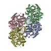
| ||||||||
|---|---|---|---|---|---|---|---|---|---|
| 1 | 
| ||||||||
| 2 | 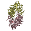
| ||||||||
| Unit cell |
|
-Components
| #1: Protein | Mass: 85544.094 Da / Num. of mol.: 4 / Mutation: C236Y Source method: isolated from a genetically manipulated source Source: (gene. exp.) Bifidobacterium longum (bacteria) / Strain: JCM1217 / Gene: BLLJ_1623, lnpA / Plasmid: pET28 / Production host: References: UniProt: E8MF13, 1,3-beta-galactosyl-N-acetylhexosamine phosphorylase #2: Sugar | ChemComp-NDG /   Type: D-saccharide, alpha linking / Mass: 221.208 Da / Num. of mol.: 4 Type: D-saccharide, alpha linking / Mass: 221.208 Da / Num. of mol.: 4Source method: isolated from a genetically manipulated source Formula: C8H15NO6 #3: Water | ChemComp-HOH / |  Mass: 18.015 Da / Num. of mol.: 900 / Source method: isolated from a natural source / Formula: H2O Mass: 18.015 Da / Num. of mol.: 900 / Source method: isolated from a natural source / Formula: H2O |
|---|
-Experimental details
-Experiment
| Experiment | Method: X-RAY DIFFRACTION / Number of used crystals: 1 |
|---|
- Sample preparation
| Crystal | Density Matthews: 2.47 Å3/Da / Density % sol: 50.3 % |
|---|---|
| Crystal grow | Temperature: 277 K / Method: vapor diffusion, sitting drop / pH: 6.5 Details: Na cacodyrate, Mg(NO3)2, PEG 4000, GlcNAc, pH 6.5, VAPOR DIFFUSION, SITTING DROP, temperature 277K |
-Data collection
| Diffraction | Mean temperature: 95 K |
|---|---|
| Diffraction source | Source: SYNCHROTRON / Site: Photon Factory  / Beamline: AR-NW12A / Wavelength: 1 Å / Beamline: AR-NW12A / Wavelength: 1 Å |
| Detector | Type: ADSC QUANTUM 210 / Detector: CCD / Date: Jun 10, 2013 |
| Radiation | Monochromator: Si 111 / Protocol: SINGLE WAVELENGTH / Monochromatic (M) / Laue (L): M / Scattering type: x-ray |
| Radiation wavelength | Wavelength: 1 Å / Relative weight: 1 |
| Reflection | Resolution: 2.6→50 Å / Num. all: 100835 / Num. obs: 97558 / % possible obs: 96.8 % / Redundancy: 2 % / Biso Wilson estimate: 28.4 Å2 / Rmerge(I) obs: 0.083 / Net I/σ(I): 10.9 |
| Reflection shell | Resolution: 2.6→2.64 Å / Redundancy: 2 % / Rmerge(I) obs: 0.213 / Mean I/σ(I) obs: 2.5 / Num. unique all: 4960 / % possible all: 97.4 |
- Processing
| Software |
| ||||||||||||||||||||||||||||||||||||||||||||||||||||||||||||||||||||||||||||||||||||||||||||||||||||||||||||||||||||||||||||||||||||||||||||||||||||||||||||||||||||||||||||||||||||||
|---|---|---|---|---|---|---|---|---|---|---|---|---|---|---|---|---|---|---|---|---|---|---|---|---|---|---|---|---|---|---|---|---|---|---|---|---|---|---|---|---|---|---|---|---|---|---|---|---|---|---|---|---|---|---|---|---|---|---|---|---|---|---|---|---|---|---|---|---|---|---|---|---|---|---|---|---|---|---|---|---|---|---|---|---|---|---|---|---|---|---|---|---|---|---|---|---|---|---|---|---|---|---|---|---|---|---|---|---|---|---|---|---|---|---|---|---|---|---|---|---|---|---|---|---|---|---|---|---|---|---|---|---|---|---|---|---|---|---|---|---|---|---|---|---|---|---|---|---|---|---|---|---|---|---|---|---|---|---|---|---|---|---|---|---|---|---|---|---|---|---|---|---|---|---|---|---|---|---|---|---|---|---|---|
| Refinement | Method to determine structure: MOLECULAR REPLACEMENT Starting model: 2zuu Resolution: 2.6→36.09 Å / Cor.coef. Fo:Fc: 0.943 / Cor.coef. Fo:Fc free: 0.887 / SU B: 10.927 / SU ML: 0.232 / Cross valid method: THROUGHOUT / ESU R Free: 0.342 / Stereochemistry target values: MAXIMUM LIKELIHOOD / Details: HYDROGENS HAVE BEEN ADDED IN THE RIDING POSITIONS
| ||||||||||||||||||||||||||||||||||||||||||||||||||||||||||||||||||||||||||||||||||||||||||||||||||||||||||||||||||||||||||||||||||||||||||||||||||||||||||||||||||||||||||||||||||||||
| Solvent computation | Ion probe radii: 0.8 Å / Shrinkage radii: 0.8 Å / VDW probe radii: 1.2 Å / Solvent model: MASK | ||||||||||||||||||||||||||||||||||||||||||||||||||||||||||||||||||||||||||||||||||||||||||||||||||||||||||||||||||||||||||||||||||||||||||||||||||||||||||||||||||||||||||||||||||||||
| Displacement parameters | Biso mean: 30.014 Å2
| ||||||||||||||||||||||||||||||||||||||||||||||||||||||||||||||||||||||||||||||||||||||||||||||||||||||||||||||||||||||||||||||||||||||||||||||||||||||||||||||||||||||||||||||||||||||
| Refinement step | Cycle: LAST / Resolution: 2.6→36.09 Å
| ||||||||||||||||||||||||||||||||||||||||||||||||||||||||||||||||||||||||||||||||||||||||||||||||||||||||||||||||||||||||||||||||||||||||||||||||||||||||||||||||||||||||||||||||||||||
| Refine LS restraints |
|