 Movie
Movie Controller

[English] 日本語
 Yorodumi
- PDB-3wbk: crystal structure analysis of eukaryotic translation initiation f... -
+ Open data

- Basic information
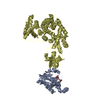
| Entry | Database: PDB / ID: 3wbk | ||||||
|---|---|---|---|---|---|---|---|
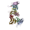
| Title | crystal structure analysis of eukaryotic translation initiation factor 5B and 1A complex | ||||||
 Components Components |
| ||||||
 Keywords Keywords | BIOSYNTHETIC PROTEIN / flexible / eukaryotic translation initiation | ||||||
| Function / homology |  Function and homology information Function and homology informationformation of translation initiation ternary complex / translation reinitiation / formation of cytoplasmic translation initiation complex / eukaryotic 43S preinitiation complex / formation of translation preinitiation complex / eukaryotic 48S preinitiation complex / protein-synthesizing GTPase / regulation of translational initiation / Formation of the ternary complex, and subsequently, the 43S complex / Translation initiation complex formation ...formation of translation initiation ternary complex / translation reinitiation / formation of cytoplasmic translation initiation complex / eukaryotic 43S preinitiation complex / formation of translation preinitiation complex / eukaryotic 48S preinitiation complex / protein-synthesizing GTPase / regulation of translational initiation / Formation of the ternary complex, and subsequently, the 43S complex / Translation initiation complex formation / Ribosomal scanning and start codon recognition / GTP hydrolysis and joining of the 60S ribosomal subunit / Formation of a pool of free 40S subunits / L13a-mediated translational silencing of Ceruloplasmin expression / ribosomal small subunit binding / translation initiation factor activity / translation initiation factor binding / cytosolic ribosome assembly / ribosome assembly / maturation of SSU-rRNA from tricistronic rRNA transcript (SSU-rRNA, 5.8S rRNA, LSU-rRNA) / translational initiation / cytoplasmic stress granule / double-stranded RNA binding / ribosome binding / small ribosomal subunit rRNA binding / cytosolic small ribosomal subunit / GTPase activity / protein kinase binding / GTP binding / mitochondrion / metal ion binding / cytoplasm / cytosol Similarity search - Function | ||||||
| Biological species |  | ||||||
| Method |  X-RAY DIFFRACTION / SYNCHROTRON / MOLECULAR REPLACEMENT / Resolution: 3.3 Å X-RAY DIFFRACTION / SYNCHROTRON / MOLECULAR REPLACEMENT / Resolution: 3.3 Å | ||||||
 Authors Authors | Zheng, A. / Yamamoto, R. / Ose, T. / Yu, J. / Tanaka, I. / Yao, M. | ||||||
 Citation Citation | Journal: Acta Crystallogr.,Sect.D / Year: 2014 Title: X-ray structures of eIF5B and the eIF5B-eIF1A complex: the conformational flexibility of eIF5B is restricted on the ribosome by interaction with eIF1A Authors: Zheng, A. / Yu, J. / Yamamoto, R. / Ose, T. / Tanaka, I. / Yao, M. | ||||||
| History |
|
- Structure visualization
| Structure viewer | Molecule: MolmilJmol/JSmol |
|---|
- Downloads & links
-Download
| PDBx/mmCIF format | 3wbk.cif.gz | 242.6 KB | Display | PDBx/mmCIF format |
|---|---|---|---|---|
| PDB format | pdb3wbk.ent.gz | 194.6 KB | Display | PDB format |
| PDBx/mmJSON format | 3wbk.json.gz | Tree view | PDBx/mmJSON format | |
| Others |  Other downloads Other downloads |
-Validation report
| Arichive directory | https://data.pdbj.org/pub/pdb/validation_reports/wb/3wbkftp://data.pdbj.org/pub/pdb/validation_reports/wb/3wbk | HTTPS FTP |
|---|
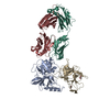
-Related structure data
| Related structure data | 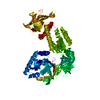 3wbiSC  3wbjC S: Starting model for refinement C: citing same article ( |
|---|---|
| Similar structure data |


-Links
 PDBj
PDBj


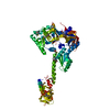
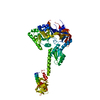
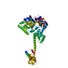
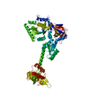
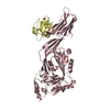
- Assembly
| Deposited unit | 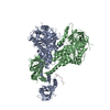
| ||||||||||||||||||
|---|---|---|---|---|---|---|---|---|---|---|---|---|---|---|---|---|---|---|---|
| 1 | 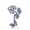
| ||||||||||||||||||
| 2 | 
| ||||||||||||||||||
| Unit cell |
| ||||||||||||||||||
| Noncrystallographic symmetry (NCS) | NCS domain:
NCS domain segments: Component-ID: _ / Ens-ID: 1 / Beg auth comp-ID: ASP / Beg label comp-ID: ASP / End auth comp-ID: GLU / End label comp-ID: GLU / Refine code: _ / Auth seq-ID: 2 - 602 / Label seq-ID: 6 - 606
|
-Components
| #1: Protein | Mass: 67724.023 Da / Num. of mol.: 2 / Fragment: UNP residues 401-1002 Source method: isolated from a genetically manipulated source Source: (gene. exp.) Strain: ATCC 204508 / S288c / Gene: FUN12, YAL035W / Production host:  #2: Protein | | Mass: 15040.341 Da / Num. of mol.: 1 / Fragment: UNP residues 27-153 Source method: isolated from a genetically manipulated source Source: (gene. exp.) Strain: ATCC 204508 / S288c / Gene: TIF11, YMR260C, YM8156.02C / Production host: Has protein modification | Y | |
|---|
-Experimental details
-Experiment
| Experiment | Method: X-RAY DIFFRACTION / Number of used crystals: 1 |
|---|
- Sample preparation
| Crystal | Density Matthews: 2.72 Å3/Da / Density % sol: 54.76 % / Mosaicity: 0.422 ° |
|---|---|
| Crystal grow | Temperature: 293 K / Method: vapor diffusion, sitting drop / pH: 8.2 Details: 100mM Tris-HCl pH 8.2, 12.5%(w/v) PEG 3350, VAPOR DIFFUSION, SITTING DROP, temperature 293K |
-Data collection
| Diffraction | Mean temperature: 100 K | |||||||||||||||||||||||||||||||||||||||||||||||||||||||||||||||||||||||||||||
|---|---|---|---|---|---|---|---|---|---|---|---|---|---|---|---|---|---|---|---|---|---|---|---|---|---|---|---|---|---|---|---|---|---|---|---|---|---|---|---|---|---|---|---|---|---|---|---|---|---|---|---|---|---|---|---|---|---|---|---|---|---|---|---|---|---|---|---|---|---|---|---|---|---|---|---|---|---|---|
| Diffraction source | Source: SYNCHROTRON / Site: SPring-8  / Beamline: BL41XU / Wavelength: 0.98 Å / Beamline: BL41XU / Wavelength: 0.98 Å | |||||||||||||||||||||||||||||||||||||||||||||||||||||||||||||||||||||||||||||
| Detector | Type: RAYONIX MX225HE / Detector: CCD / Date: Jun 28, 2010 | |||||||||||||||||||||||||||||||||||||||||||||||||||||||||||||||||||||||||||||
| Radiation | Monochromator: Rotated-inclined double-crystal monochromator , Si (111) Protocol: SINGLE WAVELENGTH / Monochromatic (M) / Laue (L): M / Scattering type: x-ray | |||||||||||||||||||||||||||||||||||||||||||||||||||||||||||||||||||||||||||||
| Radiation wavelength | Wavelength: 0.98 Å / Relative weight: 1 | |||||||||||||||||||||||||||||||||||||||||||||||||||||||||||||||||||||||||||||
| Reflection | Resolution: 3.3→50 Å / Num. obs: 24875 / % possible obs: 98.5 % / Observed criterion σ(I): -1.15 / Redundancy: 9.4 % / Rmerge(I) obs: 0.071 / Χ2: 1.371 / Net I/σ(I): 16.2 | |||||||||||||||||||||||||||||||||||||||||||||||||||||||||||||||||||||||||||||
| Reflection shell |
|
- Processing
| Software |
| ||||||||||||||||||||||||||||||||||||||||||||||||||||||||||||
|---|---|---|---|---|---|---|---|---|---|---|---|---|---|---|---|---|---|---|---|---|---|---|---|---|---|---|---|---|---|---|---|---|---|---|---|---|---|---|---|---|---|---|---|---|---|---|---|---|---|---|---|---|---|---|---|---|---|---|---|---|---|
| Refinement | Method to determine structure: MOLECULAR REPLACEMENT Starting model: PDB ENTRY 3WBI Resolution: 3.3→40 Å / Cor.coef. Fo:Fc: 0.926 / Cor.coef. Fo:Fc free: 0.89 / WRfactor Rfree: 0.3272 / WRfactor Rwork: 0.2706 / Occupancy max: 1 / Occupancy min: 1 / FOM work R set: 0.7357 / SU B: 36.687 / SU ML: 0.608 / SU Rfree: 0.6868 / Cross valid method: THROUGHOUT / σ(F): 0 / ESU R Free: 0.687 / Stereochemistry target values: MAXIMUM LIKELIHOOD Details: HYDROGENS HAVE BEEN USED IF PRESENT IN THE INPUT U VALUES: REFINED INDIVIDUALLY
| ||||||||||||||||||||||||||||||||||||||||||||||||||||||||||||
| Solvent computation | Ion probe radii: 0.8 Å / Shrinkage radii: 0.8 Å / VDW probe radii: 1.2 Å / Solvent model: BABINET MODEL WITH MASK | ||||||||||||||||||||||||||||||||||||||||||||||||||||||||||||
| Displacement parameters | Biso max: 349.8 Å2 / Biso mean: 159.3338 Å2 / Biso min: 47.82 Å2
| ||||||||||||||||||||||||||||||||||||||||||||||||||||||||||||
| Refinement step | Cycle: LAST / Resolution: 3.3→40 Å
| ||||||||||||||||||||||||||||||||||||||||||||||||||||||||||||
| Refine LS restraints |
| ||||||||||||||||||||||||||||||||||||||||||||||||||||||||||||
| Refine LS restraints NCS | Ens-ID: 1 / Number: 626 / Refine-ID: X-RAY DIFFRACTION / Type: interatomic distance / Rms dev position: 0.23 Å / Weight position: 0.05
| ||||||||||||||||||||||||||||||||||||||||||||||||||||||||||||
| LS refinement shell | Resolution: 3.304→3.389 Å / Total num. of bins used: 20
|
