 Movie
Movie Controller

[English] 日本語
 Yorodumi
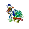
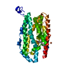
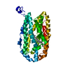
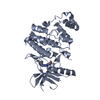
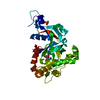
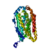
- PDB-3w6z: Crystal structure of NADP bound L-serine 3-dehydrogenase (K170M) ... -
+ Open data

- Basic information
| Entry | Database: PDB / ID: 3w6z | ||||||
|---|---|---|---|---|---|---|---|
| Title | Crystal structure of NADP bound L-serine 3-dehydrogenase (K170M) from Hyperthermophilic Archaeon Pyrobaculum calidifontis | ||||||
 Components Components | 6-phosphogluconate dehydrogenase, NAD-binding protein | ||||||
 Keywords Keywords | OXIDOREDUCTASE / HYPERTHERMOPHILIC ARCHAEON / ROSSMANN FOLD / L-SERINE 3-DEHYDROGENASE / NAD(P) BINDING | ||||||
| Function / homology |  Function and homology information Function and homology informationL-serine 3-dehydrogenase (NAD+) / NAD binding / NADP binding / oxidoreductase activity Similarity search - Function | ||||||
| Biological species |   Pyrobaculum calidifontis (archaea) Pyrobaculum calidifontis (archaea) | ||||||
| Method |  X-RAY DIFFRACTION / SYNCHROTRON / MOLECULAR REPLACEMENT / Resolution: 1.44 Å X-RAY DIFFRACTION / SYNCHROTRON / MOLECULAR REPLACEMENT / Resolution: 1.44 Å | ||||||
 Authors Authors | Yoneda, K. / Sakuraba, H. / Ohshima, T. | ||||||
 Citation Citation | Journal: Extremophiles / Year: 2018 Title: Crystal structure of the NADP+and tartrate-bound complex of L-serine 3-dehydrogenase from the hyperthermophilic archaeon Pyrobaculum calidifontis. Authors: Yoneda, K. / Sakuraba, H. / Araki, T. / Ohshima, T. | ||||||
| History |
|
- Structure visualization
| Structure viewer | Molecule: MolmilJmol/JSmol |
|---|
- Downloads & links
-Download
| PDBx/mmCIF format | 3w6z.cif.gz | 75.5 KB | Display | PDBx/mmCIF format |
|---|---|---|---|---|
| PDB format | pdb3w6z.ent.gz | 54.6 KB | Display | PDB format |
| PDBx/mmJSON format | 3w6z.json.gz | Tree view | PDBx/mmJSON format | |
| Others |  Other downloads Other downloads |
-Validation report
| Arichive directory | https://data.pdbj.org/pub/pdb/validation_reports/w6/3w6zftp://data.pdbj.org/pub/pdb/validation_reports/w6/3w6z | HTTPS FTP |
|---|
-Related structure data
| Related structure data |  3w6uSC  3ws7C  5xvhC S: Starting model for refinement C: citing same article ( |
|---|---|
| Similar structure data |


-Links
 PDBj
PDBj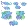
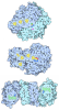
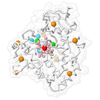
- Assembly
| Deposited unit | 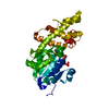
| ||||||||
|---|---|---|---|---|---|---|---|---|---|
| 1 |
| ||||||||
| 2 | 
| ||||||||
| Unit cell |
| ||||||||
| Components on special symmetry positions |
|
-Components
| #1: Protein | Mass: 32835.266 Da / Num. of mol.: 1 / Mutation: K170M Source method: isolated from a genetically manipulated source Source: (gene. exp.) Pyrobaculum calidifontis (archaea) / Strain: JCM 11548 / VA1 / Gene: Pcal_0699 / Plasmid: pET15b / Production host:   Escherichia coli (E. coli) / References: UniProt: A3MU08, EC: 1.1.1.276 Escherichia coli (E. coli) / References: UniProt: A3MU08, EC: 1.1.1.276 |
|---|---|
| #2: Chemical | ChemComp-NAP /  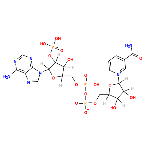 Mass: 743.405 Da / Num. of mol.: 1 / Source method: obtained synthetically / Formula: C21H28N7O17P3 Mass: 743.405 Da / Num. of mol.: 1 / Source method: obtained synthetically / Formula: C21H28N7O17P3 |
| #3: Water | ChemComp-HOH /  Mass: 18.015 Da / Num. of mol.: 198 / Source method: isolated from a natural source / Formula: H2O Mass: 18.015 Da / Num. of mol.: 198 / Source method: isolated from a natural source / Formula: H2O |
-Experimental details
-Experiment
| Experiment | Method: X-RAY DIFFRACTION / Number of used crystals: 1 |
|---|
- Sample preparation
| Crystal | Density Matthews: 2.85 Å3/Da / Density % sol: 56.89 % |
|---|---|
| Crystal grow | Temperature: 293 K / Method: vapor diffusion, sitting drop / pH: 4.5 Details: 2.0M AMMONIUM SULFATE, 0.1M ACETATE BUFFER, pH 4.5, VAPOR DIFFUSION, SITTING DROP, temperature 293K |
-Data collection
| Diffraction | Mean temperature: 100 K |
|---|---|
| Diffraction source | Source: SYNCHROTRON / Site: Photon Factory  / Beamline: BL-1A / Wavelength: 1.1 Å / Beamline: BL-1A / Wavelength: 1.1 Å |
| Detector | Type: PILATUS 2M-F / Detector: CCD / Date: Oct 30, 2012 |
| Radiation | Protocol: SINGLE WAVELENGTH / Monochromatic (M) / Laue (L): M / Scattering type: x-ray |
| Radiation wavelength | Wavelength: 1.1 Å / Relative weight: 1 |
| Reflection | Resolution: 1.44→57.938 Å / Num. obs: 67330 / Biso Wilson estimate: 16.4 Å2 |
- Processing
| Software |
| ||||||||||||||||||
|---|---|---|---|---|---|---|---|---|---|---|---|---|---|---|---|---|---|---|---|
| Refinement | Method to determine structure: MOLECULAR REPLACEMENT Starting model: 3W6U Resolution: 1.44→46.8 Å /
| ||||||||||||||||||
| Refinement step | Cycle: LAST / Resolution: 1.44→46.8 Å
|
