 Movie
Movie Controller

[English] 日本語
 Yorodumi
- PDB-3w01: Crystal structure of PcrB complexed with PEG from Staphylococcus ... -
+ Open data

- Basic information
| Entry | Database: PDB / ID: 3w01 | ||||||
|---|---|---|---|---|---|---|---|
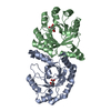
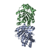
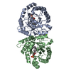
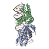
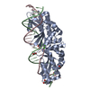
| Title | Crystal structure of PcrB complexed with PEG from Staphylococcus aureus subsp. aureus Mu3 | ||||||
 Components Components | Heptaprenylglyceryl phosphate synthase | ||||||
 Keywords Keywords | TRANSFERASE / biosynthesis / prenyltransferases / enzyme catalysis | ||||||
| Function / homology |  Function and homology information Function and homology information: / heptaprenylglyceryl phosphate synthase activity / glycerophospholipid biosynthetic process / magnesium ion binding Similarity search - Function | ||||||
| Biological species |   Staphylococcus aureus (bacteria) Staphylococcus aureus (bacteria) | ||||||
| Method |  X-RAY DIFFRACTION / SYNCHROTRON / MOLECULAR REPLACEMENT / Resolution: 1.54 Å X-RAY DIFFRACTION / SYNCHROTRON / MOLECULAR REPLACEMENT / Resolution: 1.54 Å | ||||||
 Authors Authors | Ren, F. / Feng, X. / Ko, T.P. / Huang, C.H. / Hu, Y. / Chan, H.C. / Liu, Y.L. / Wang, K. / Chen, C.C. / Pang, X. ...Ren, F. / Feng, X. / Ko, T.P. / Huang, C.H. / Hu, Y. / Chan, H.C. / Liu, Y.L. / Wang, K. / Chen, C.C. / Pang, X. / He, M. / Li, Y. / Oldfield, E. / Guo, R.T. | ||||||
 Citation Citation | Journal: Chembiochem / Year: 2013 Title: Insights into TIM-barrel prenyl transferase mechanisms: crystal structures of PcrB from Bacillus subtilis and Staphylococcus aureus Authors: Ren, F. / Feng, X. / Ko, T.P. / Huang, C.H. / Hu, Y. / Chan, H.C. / Liu, Y.L. / Wang, K. / Chen, C.C. / Pang, X. / He, M. / Li, Y. / Oldfield, E. / Guo, R.T. | ||||||
| History |
|
- Structure visualization
| Structure viewer | Molecule: MolmilJmol/JSmol |
|---|
- Downloads & links
-Download
| PDBx/mmCIF format | 3w01.cif.gz | 115 KB | Display | PDBx/mmCIF format |
|---|---|---|---|---|
| PDB format | pdb3w01.ent.gz | 87.7 KB | Display | PDB format |
| PDBx/mmJSON format | 3w01.json.gz | Tree view | PDBx/mmJSON format | |
| Others |  Other downloads Other downloads |
-Validation report
| Arichive directory | https://data.pdbj.org/pub/pdb/validation_reports/w0/3w01ftp://data.pdbj.org/pub/pdb/validation_reports/w0/3w01 | HTTPS FTP |
|---|
-Related structure data
| Related structure data |  3vzxSC  3vzyC  3vzzC  3w00C  3w02C S: Starting model for refinement C: citing same article ( |
|---|---|
| Similar structure data |


-Links
 PDBj
PDBj- Assembly
| Deposited unit | 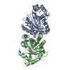
| ||||||||
|---|---|---|---|---|---|---|---|---|---|
| 1 |
| ||||||||
| Unit cell |
|
-Components
| #1: Protein | Mass: 26202.826 Da / Num. of mol.: 2 Source method: isolated from a genetically manipulated source Source: (gene. exp.) Staphylococcus aureus (bacteria) / Strain: Mu3 / Gene: pcrB / Plasmid: pET32a / Production host: References: UniProt: A7X435, Transferases; Transferring alkyl or aryl groups, other than methyl groups #2: Chemical | ChemComp-PGE / |   Mass: 150.173 Da / Num. of mol.: 1 / Source method: obtained synthetically / Formula: C6H14O4 Mass: 150.173 Da / Num. of mol.: 1 / Source method: obtained synthetically / Formula: C6H14O4#3: Water | ChemComp-HOH / |  Mass: 18.015 Da / Num. of mol.: 608 / Source method: isolated from a natural source / Formula: H2O Mass: 18.015 Da / Num. of mol.: 608 / Source method: isolated from a natural source / Formula: H2O |
|---|
-Experimental details
-Experiment
| Experiment | Method: X-RAY DIFFRACTION / Number of used crystals: 1 |
|---|
- Sample preparation
| Crystal | Density Matthews: 2.16 Å3/Da / Density % sol: 43.05 % |
|---|---|
| Crystal grow | Temperature: 295 K / Method: vapor diffusion, sitting drop / pH: 8.5 Details: 0.1M Tris, 21% PEG3350, pH 8.5, VAPOR DIFFUSION, SITTING DROP, temperature 295K |
-Data collection
| Diffraction | Mean temperature: 100 K |
|---|---|
| Diffraction source | Source: SYNCHROTRON / Site: NSRRC  / Beamline: BL13B1 / Wavelength: 1 Å / Beamline: BL13B1 / Wavelength: 1 Å |
| Detector | Type: ADSC QUANTUM 315 / Detector: CCD / Date: Feb 18, 2012 |
| Radiation | Protocol: SINGLE WAVELENGTH / Monochromatic (M) / Laue (L): M / Scattering type: x-ray |
| Radiation wavelength | Wavelength: 1 Å / Relative weight: 1 |
| Reflection | Resolution: 1.54→25 Å / Num. obs: 66696 / % possible obs: 98.1 % / Redundancy: 7.6 % / Rmerge(I) obs: 0.053 / Net I/σ(I): 44.8 |
| Reflection shell | Resolution: 1.54→1.6 Å / Redundancy: 5.2 % / Rmerge(I) obs: 0.449 / Mean I/σ(I) obs: 3.7 / Num. unique all: 5680 / % possible all: 84.5 |
- Processing
| Software |
| ||||||||||||||||||||
|---|---|---|---|---|---|---|---|---|---|---|---|---|---|---|---|---|---|---|---|---|---|
| Refinement | Method to determine structure: MOLECULAR REPLACEMENT Starting model: 3VZX Resolution: 1.54→24.9 Å / σ(F): 2 / Stereochemistry target values: Engh & Huber
| ||||||||||||||||||||
| Refinement step | Cycle: LAST / Resolution: 1.54→24.9 Å
| ||||||||||||||||||||
| LS refinement shell | Resolution: 1.54→1.6 Å
|
