 Movie
Movie Controller


+ Open data

- Basic information
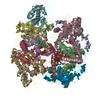
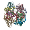
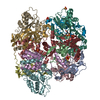
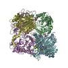
| Entry | Database: PDB / ID: 3vyg | ||||||
|---|---|---|---|---|---|---|---|
| Title | Crystal structure of Thiocyanate hydrolase mutant R136W | ||||||
 Components Components | (Thiocyanate hydrolase subunit ...) x 3 | ||||||
 Keywords Keywords | HYDROLASE / Thiocyanate hydrolase / Cysteine-sulfenic acid / Cysteine-sulfic acid / metalloenzyme | ||||||
| Function / homology |  Function and homology information Function and homology informationthiocyanate hydrolase / thiocyanate hydrolase activity / thiocyanate catabolic process / transition metal ion binding Similarity search - Function | ||||||
| Biological species |  Thiobacillus thioparus (bacteria) Thiobacillus thioparus (bacteria) | ||||||
| Method |  X-RAY DIFFRACTION / SYNCHROTRON / MOLECULAR REPLACEMENT / Resolution: 1.72 Å X-RAY DIFFRACTION / SYNCHROTRON / MOLECULAR REPLACEMENT / Resolution: 1.72 Å | ||||||
 Authors Authors | Yamanaka, Y. / Sato, M. / Arakawa, T. / Namima, S. / Hori, S. / Ohtaki, A. / Noguchi, K. / Katayama, Y. / Yohda, M. / Odaka, M. | ||||||
 Citation Citation | Journal: To be published Title: Effects of argnine residue around the substrate pocket on the substrate specificity of thiocyanate hydrolase Authors: Yamanaka, Y. / Sato, M. / Arakawa, T. / Namima, S. / Hori, S. / Ohtaki, A. / Noguchi, K. / Katayama, Y. / Yohda, M. / Odaka, M. | ||||||
| History |
|
- Structure visualization
| Structure viewer | Molecule: MolmilJmol/JSmol |
|---|
- Downloads & links
-Download
| PDBx/mmCIF format | 3vyg.cif.gz | 427.5 KB | Display | PDBx/mmCIF format |
|---|---|---|---|---|
| PDB format | pdb3vyg.ent.gz | 346.5 KB | Display | PDB format |
| PDBx/mmJSON format | 3vyg.json.gz | Tree view | PDBx/mmJSON format | |
| Others |  Other downloads Other downloads |
-Validation report
| Arichive directory | https://data.pdbj.org/pub/pdb/validation_reports/vy/3vygftp://data.pdbj.org/pub/pdb/validation_reports/vy/3vyg | HTTPS FTP |
|---|
-Related structure data
| Related structure data | 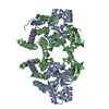 3vyhC  2dd5S C: citing same article ( S: Starting model for refinement |
|---|---|
| Similar structure data |


-Links
 PDBj
PDBj


- Assembly
| Deposited unit | 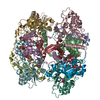
| ||||||||
|---|---|---|---|---|---|---|---|---|---|
| 1 |
| ||||||||
| Unit cell |
|
-Components
-Thiocyanate hydrolase subunit ... , 3 types, 12 molecules ADGJBEHKCFIL
| #1: Protein | Mass: 14507.152 Da / Num. of mol.: 4 Source method: isolated from a genetically manipulated source Source: (gene. exp.) Thiobacillus thioparus (bacteria) / Gene: scnA / Production host: #2: Protein | Mass: 18065.549 Da / Num. of mol.: 4 Source method: isolated from a genetically manipulated source Source: (gene. exp.) Thiobacillus thioparus (bacteria) / Gene: scnB / Production host: #3: Protein | Mass: 27648.445 Da / Num. of mol.: 4 / Mutation: R136W Source method: isolated from a genetically manipulated source Source: (gene. exp.) Thiobacillus thioparus (bacteria) / Gene: scnC / Production host: |
|---|
-Non-polymers , 3 types, 1508 molecules 


| #4: Chemical | ChemComp-3CO /  Mass: 58.933 Da / Num. of mol.: 4 / Source method: obtained synthetically / Formula: Co Mass: 58.933 Da / Num. of mol.: 4 / Source method: obtained synthetically / Formula: Co#5: Chemical | ChemComp-TLA /  Mass: 150.087 Da / Num. of mol.: 4 / Source method: obtained synthetically / Formula: C4H6O6 Mass: 150.087 Da / Num. of mol.: 4 / Source method: obtained synthetically / Formula: C4H6O6#6: Water | ChemComp-HOH / | Mass: 18.015 Da / Num. of mol.: 1500 / Source method: isolated from a natural source / Formula: H2O |
|---|
-Details
| Has protein modification | Y |
|---|
-Experimental details
-Experiment
| Experiment | Method: X-RAY DIFFRACTION / Number of used crystals: 1 |
|---|
- Sample preparation
| Crystal | Density Matthews: 3.5 Å3/Da / Density % sol: 64.88 % |
|---|---|
| Crystal grow | Temperature: 293 K / Method: vapor diffusion, hanging drop / pH: 6.8 Details: 0.1M potassium phosphate pH 6.8, 1.4M potassium phosphate tartrate, VAPOR DIFFUSION, HANGING DROP, temperature 293K |
-Data collection
| Diffraction | Mean temperature: 95 K |
|---|---|
| Diffraction source | Source: SYNCHROTRON / Site: Photon Factory  / Beamline: AR-NW12A / Wavelength: 1 Å / Beamline: AR-NW12A / Wavelength: 1 Å |
| Detector | Type: ADSC QUANTUM 210r / Detector: CCD / Date: Dec 17, 2011 |
| Radiation | Monochromator: Si 111 CHANNEL / Protocol: SINGLE WAVELENGTH / Monochromatic (M) / Laue (L): M / Scattering type: x-ray |
| Radiation wavelength | Wavelength: 1 Å / Relative weight: 1 |
| Reflection | Resolution: 1.72→50 Å / Num. all: 343322 / Num. obs: 325674 / % possible obs: 96.5 % / Rmerge(I) obs: 0.062 |
| Reflection shell | Resolution: 1.72→1.78 Å / Rmerge(I) obs: 0.256 / Mean I/σ(I) obs: 9.2 / % possible all: 99.8 |
- Processing
| Software |
| |||||||||||||||||||||||||||||||||||||||||||||||||||||||||||||||||
|---|---|---|---|---|---|---|---|---|---|---|---|---|---|---|---|---|---|---|---|---|---|---|---|---|---|---|---|---|---|---|---|---|---|---|---|---|---|---|---|---|---|---|---|---|---|---|---|---|---|---|---|---|---|---|---|---|---|---|---|---|---|---|---|---|---|---|
| Refinement | Method to determine structure: MOLECULAR REPLACEMENT Starting model: 2DD5 Resolution: 1.72→33.96 Å / Cor.coef. Fo:Fc: 0.943 / Cor.coef. Fo:Fc free: 0.927 / SU B: 1.626 / SU ML: 0.054 / Cross valid method: THROUGHOUT / ESU R: 0.086 / ESU R Free: 0.089 / Stereochemistry target values: MAXIMUM LIKELIHOOD / Details: HYDROGENS HAVE BEEN ADDED IN THE RIDING POSITIONS
| |||||||||||||||||||||||||||||||||||||||||||||||||||||||||||||||||
| Solvent computation | Ion probe radii: 0.8 Å / Shrinkage radii: 0.8 Å / VDW probe radii: 1.4 Å / Solvent model: MASK | |||||||||||||||||||||||||||||||||||||||||||||||||||||||||||||||||
| Displacement parameters | Biso mean: 20.084 Å2
| |||||||||||||||||||||||||||||||||||||||||||||||||||||||||||||||||
| Refinement step | Cycle: LAST / Resolution: 1.72→33.96 Å
| |||||||||||||||||||||||||||||||||||||||||||||||||||||||||||||||||
| Refine LS restraints |
| |||||||||||||||||||||||||||||||||||||||||||||||||||||||||||||||||
| LS refinement shell | Resolution: 1.721→1.766 Å / Total num. of bins used: 20
|
