 Movie
Movie Controller

[English] 日本語
 Yorodumi
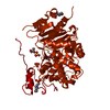
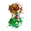
- PDB-3vwr: Crystal structure of 6-aminohexanoate-dimer hydrolase S112A/G181D... -
+ Open data

- Basic information
| Entry | Database: PDB / ID: 3vwr | |||||||||
|---|---|---|---|---|---|---|---|---|---|---|
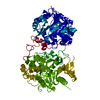
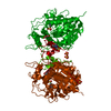
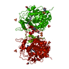
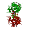
| Title | Crystal structure of 6-aminohexanoate-dimer hydrolase S112A/G181D/R187G/H266N/D370Y mutant complexd with 6-aminohexanoate | |||||||||
 Components Components | 6-aminohexanoate-dimer hydrolase | |||||||||
 Keywords Keywords | HYDROLASE / NYLON DEGRADATION | |||||||||
| Function / homology |  Function and homology information Function and homology information6-aminohexanoate-oligomer exohydrolase / 6-aminohexanoate-dimer hydrolase activity / nylon catabolic process Similarity search - Function | |||||||||
| Biological species |  Flavobacterium (bacteria) Flavobacterium (bacteria) | |||||||||
| Method |  X-RAY DIFFRACTION / FOURIER SYNTHESIS / Resolution: 1.65 Å X-RAY DIFFRACTION / FOURIER SYNTHESIS / Resolution: 1.65 Å | |||||||||
 Authors Authors | Kawashima, Y. / Shibata, N. / Negoro, S. / Higuchi, Y. | |||||||||
 Citation Citation | Journal: To be Published Title: Structural, kinetic and theoretical analyses of hydrolase mutants altering in the directionality and equilibrium point of reversible amide-synthetic/hydrolytic reaction Authors: Negoro, S. / Kawashima, Y. / Shibata, N. / Shigeta, Y. / Kobayashi, T. / Nishiguchi, H. / Matsui, T. / Baba, T. / Lee, Y. / Kamiya, K. / Kato, D. / Takeo, M. / Higuchi, Y. | |||||||||
| History |
|
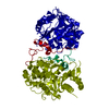
- Structure visualization
| Structure viewer | Molecule: MolmilJmol/JSmol |
|---|
- Downloads & links
-Download
| PDBx/mmCIF format | 3vwr.cif.gz | 99.9 KB | Display | PDBx/mmCIF format |
|---|---|---|---|---|
| PDB format | pdb3vwr.ent.gz | 73.6 KB | Display | PDB format |
| PDBx/mmJSON format | 3vwr.json.gz | Tree view | PDBx/mmJSON format | |
| Others |  Other downloads Other downloads |
-Validation report
| Arichive directory | https://data.pdbj.org/pub/pdb/validation_reports/vw/3vwrftp://data.pdbj.org/pub/pdb/validation_reports/vw/3vwr | HTTPS FTP |
|---|
-Related structure data


-Links
 PDBj
PDBj

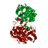
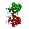
- Assembly
| Deposited unit | 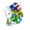
| |||||||||
|---|---|---|---|---|---|---|---|---|---|---|
| 1 | 
| |||||||||
| Unit cell |
| |||||||||
| Components on special symmetry positions |
|
-Components
-Protein , 1 types, 1 molecules A
| #1: Protein | Mass: 42805.434 Da / Num. of mol.: 1 / Mutation: S112A, G181D, R187G, H266N, D370Y Source method: isolated from a genetically manipulated source Details: THE FUSION PROTEIN OF RESIDUES 1-21 FROM NYLON OLIGOMERS-DEGRADING ENZYME EII (UNP P07061) AND RESIDUES 22-392 FROM NYLON OLIGOMERS-DEGRADING ENZYME EII' (UNP Q59710) Source: (gene. exp.) Flavobacterium (bacteria) / Strain: K172, KI723T1 / Gene: nylB, nylB' / Plasmid: PKP1500 / Production host: References: UniProt: P07061, UniProt: Q59710, UniProt: P07062*PLUS, 6-aminohexanoate-oligomer exohydrolase |
|---|
-Non-polymers , 5 types, 391 molecules 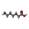


| #2: Chemical | ChemComp-ACA /  Type: peptide linking / Mass: 131.173 Da / Num. of mol.: 1 / Source method: obtained synthetically / Formula: C6H13NO2 Type: peptide linking / Mass: 131.173 Da / Num. of mol.: 1 / Source method: obtained synthetically / Formula: C6H13NO2 | ||||||
|---|---|---|---|---|---|---|---|
| #3: Chemical | ChemComp-GOL /  Mass: 92.094 Da / Num. of mol.: 5 / Source method: obtained synthetically / Formula: C3H8O3 Mass: 92.094 Da / Num. of mol.: 5 / Source method: obtained synthetically / Formula: C3H8O3#4: Chemical |  Mass: 195.237 Da / Num. of mol.: 2 / Source method: obtained synthetically / Formula: C6H13NO4S / Comment: pH buffer*YM Mass: 195.237 Da / Num. of mol.: 2 / Source method: obtained synthetically / Formula: C6H13NO4S / Comment: pH buffer*YM#5: Chemical | ChemComp-SO4 / |  Mass: 96.063 Da / Num. of mol.: 1 / Source method: obtained synthetically / Formula: SO4 Mass: 96.063 Da / Num. of mol.: 1 / Source method: obtained synthetically / Formula: SO4#6: Water | ChemComp-HOH / | Mass: 18.015 Da / Num. of mol.: 382 / Source method: isolated from a natural source / Formula: H2O |
-Experimental details
-Experiment
| Experiment | Method: X-RAY DIFFRACTION / Number of used crystals: 1 |
|---|
- Sample preparation
| Crystal | Density Matthews: 3.53 Å3/Da / Density % sol: 65.19 % / Mosaicity: 0.3 ° |
|---|---|
| Crystal grow | Temperature: 283 K / Method: vapor diffusion, sitting drop / pH: 6.5 Details: 2.2M AMMONIUM SULFATE, 0.2M LITHIUM SULFATE, 0.1M MES, pH 6.5, vapor diffusion, sitting drop, temperature 283K |
-Data collection
| Diffraction | Mean temperature: 100 K | |||||||||||||||||||||||||||||||||||||||||||||||||||||||||||||||||||||||||||||||||||||||||||||||||||
|---|---|---|---|---|---|---|---|---|---|---|---|---|---|---|---|---|---|---|---|---|---|---|---|---|---|---|---|---|---|---|---|---|---|---|---|---|---|---|---|---|---|---|---|---|---|---|---|---|---|---|---|---|---|---|---|---|---|---|---|---|---|---|---|---|---|---|---|---|---|---|---|---|---|---|---|---|---|---|---|---|---|---|---|---|---|---|---|---|---|---|---|---|---|---|---|---|---|---|---|---|
| Diffraction source | Source: ROTATING ANODE / Type: RIGAKU MICROMAX-007 / Wavelength: 1.5418 Å | |||||||||||||||||||||||||||||||||||||||||||||||||||||||||||||||||||||||||||||||||||||||||||||||||||
| Detector | Type: RIGAKU RAXIS VII / Detector: IMAGE PLATE / Date: Aug 26, 2009 | |||||||||||||||||||||||||||||||||||||||||||||||||||||||||||||||||||||||||||||||||||||||||||||||||||
| Radiation | Monochromator: CONFOCAL MIRROR / Protocol: SINGLE WAVELENGTH / Monochromatic (M) / Laue (L): M / Scattering type: x-ray | |||||||||||||||||||||||||||||||||||||||||||||||||||||||||||||||||||||||||||||||||||||||||||||||||||
| Radiation wavelength | Wavelength: 1.5418 Å / Relative weight: 1 | |||||||||||||||||||||||||||||||||||||||||||||||||||||||||||||||||||||||||||||||||||||||||||||||||||
| Reflection | Resolution: 1.6→30.38 Å / Num. obs: 80141 / % possible obs: 99.1 % / Redundancy: 4.82 % / Rmerge(I) obs: 0.032 / Χ2: 0.79 / Net I/σ(I): 21.8 / Scaling rejects: 2923 | |||||||||||||||||||||||||||||||||||||||||||||||||||||||||||||||||||||||||||||||||||||||||||||||||||
| Reflection shell | Diffraction-ID: 1
|
- Processing
| Software |
| ||||||||||||||||||||||||||||||||||||
|---|---|---|---|---|---|---|---|---|---|---|---|---|---|---|---|---|---|---|---|---|---|---|---|---|---|---|---|---|---|---|---|---|---|---|---|---|---|
| Refinement | Method to determine structure: FOURIER SYNTHESIS / Resolution: 1.65→29.65 Å / Rfactor Rfree error: 0.002 / Occupancy max: 1 / Occupancy min: 0.5 / Data cutoff high absF: 719327 / Data cutoff low absF: 0 / Isotropic thermal model: RESTRAINED / Cross valid method: THROUGHOUT / σ(F): 0 / Details: BULK SOLVENT MODEL USED
| ||||||||||||||||||||||||||||||||||||
| Solvent computation | Solvent model: FLAT MODEL / Bsol: 60.6151 Å2 / ksol: 0.45 e/Å3 | ||||||||||||||||||||||||||||||||||||
| Displacement parameters | Biso max: 76.8 Å2 / Biso mean: 21.9393 Å2 / Biso min: 6.44 Å2
| ||||||||||||||||||||||||||||||||||||
| Refine analyze |
| ||||||||||||||||||||||||||||||||||||
| Refinement step | Cycle: LAST / Resolution: 1.65→29.65 Å
| ||||||||||||||||||||||||||||||||||||
| Refine LS restraints |
| ||||||||||||||||||||||||||||||||||||
| LS refinement shell | Resolution: 1.65→1.71 Å / Rfactor Rfree error: 0.01 / Total num. of bins used: 10
| ||||||||||||||||||||||||||||||||||||
| Xplor file |
|
