 Movie
Movie Controller

[English] 日本語
 Yorodumi
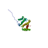
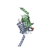
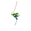
- PDB-3vuz: Crystal structure of histone methyltransferase SET7/9 in complex ... -
+ Open data

- Basic information
| Entry | Database: PDB / ID: 3vuz | ||||||
|---|---|---|---|---|---|---|---|
| Title | Crystal structure of histone methyltransferase SET7/9 in complex with AAM-1 | ||||||
 Components Components | Histone-lysine N-methyltransferase SETD7 | ||||||
 Keywords Keywords | TRANSFERASE/TRANSFERASE INHIBITOR / SET domain / transferase / adenosylmethionine binding / TRANSFERASE-TRANSFERASE INHIBITOR complex | ||||||
| Function / homology |  Function and homology information Function and homology informationpeptidyl-lysine monomethylation / peptidyl-lysine dimethylation / heterochromatin organization / [histone H3]-lysine4 N-methyltransferase / histone H3K4 monomethyltransferase activity / protein-lysine N-methyltransferase activity / histone H3 methyltransferase activity / histone methyltransferase activity / negative regulation of DNA repair / PKMTs methylate histone lysines ...peptidyl-lysine monomethylation / peptidyl-lysine dimethylation / heterochromatin organization / [histone H3]-lysine4 N-methyltransferase / histone H3K4 monomethyltransferase activity / protein-lysine N-methyltransferase activity / histone H3 methyltransferase activity / histone methyltransferase activity / negative regulation of DNA repair / PKMTs methylate histone lysines / p53 binding / chromosome / chromatin organization / response to ethanol / DNA repair / chromatin binding / DNA damage response / nucleolus / positive regulation of DNA-templated transcription / nucleoplasm / nucleus Similarity search - Function | ||||||
| Biological species |  Homo sapiens (human) Homo sapiens (human) | ||||||
| Method |  X-RAY DIFFRACTION / SYNCHROTRON / MOLECULAR REPLACEMENT / Resolution: 2.5 Å X-RAY DIFFRACTION / SYNCHROTRON / MOLECULAR REPLACEMENT / Resolution: 2.5 Å | ||||||
 Authors Authors | Niwa, H. / Handa, N. / Tomabechi, Y. / Honda, K. / Toyama, M. / Ohsawa, N. / Shirouzu, M. / Kagechika, H. / Hirano, T. / Umehara, T. / Yokoyama, S. | ||||||
 Citation Citation | Journal: Acta Crystallogr.,Sect.D / Year: 2013 Title: Structures of histone methyltransferase SET7/9 in complexes with adenosylmethionine derivatives Authors: Niwa, H. / Handa, N. / Tomabechi, Y. / Honda, K. / Toyama, M. / Ohsawa, N. / Shirouzu, M. / Kagechika, H. / Hirano, T. / Umehara, T. / Yokoyama, S. | ||||||
| History |
|
- Structure visualization
| Structure viewer | Molecule: MolmilJmol/JSmol |
|---|
- Downloads & links
-Download
| PDBx/mmCIF format | 3vuz.cif.gz | 115 KB | Display | PDBx/mmCIF format |
|---|---|---|---|---|
| PDB format | pdb3vuz.ent.gz | 87.3 KB | Display | PDB format |
| PDBx/mmJSON format | 3vuz.json.gz | Tree view | PDBx/mmJSON format | |
| Others |  Other downloads Other downloads |
-Validation report
| Arichive directory | https://data.pdbj.org/pub/pdb/validation_reports/vu/3vuzftp://data.pdbj.org/pub/pdb/validation_reports/vu/3vuz | HTTPS FTP |
|---|
-Related structure data
| Related structure data |  3vv0C 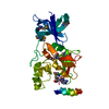 1n6aS C: citing same article ( S: Starting model for refinement |
|---|---|
| Similar structure data |


-Links
 PDBj
PDBj- Assembly
| Deposited unit | 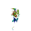
| ||||||||
|---|---|---|---|---|---|---|---|---|---|
| 1 |
| ||||||||
| Unit cell |
|
-Components
| #1: Protein | Mass: 29083.244 Da / Num. of mol.: 1 / Fragment: UNP residues 111-366 Source method: isolated from a genetically manipulated source Source: (gene. exp.) Homo sapiens (human) / Gene: SETD7, KIAA1717, KMT7, SET7, SET9 / Plasmid: PX100709-08 / Production host: CELL-FREE SYNTHESIS (others)References: UniProt: Q8WTS6, histone-lysine N-methyltransferase |
|---|---|
| #2: Chemical | ChemComp-K15 /   Mass: 451.520 Da / Num. of mol.: 1 / Source method: obtained synthetically / Formula: C20H33N7O5 Mass: 451.520 Da / Num. of mol.: 1 / Source method: obtained synthetically / Formula: C20H33N7O5 |
| #3: Water | ChemComp-HOH /  Mass: 18.015 Da / Num. of mol.: 69 / Source method: isolated from a natural source / Formula: H2O Mass: 18.015 Da / Num. of mol.: 69 / Source method: isolated from a natural source / Formula: H2O |
-Experimental details
-Experiment
| Experiment | Method: X-RAY DIFFRACTION / Number of used crystals: 1 |
|---|
- Sample preparation
| Crystal | Density Matthews: 2.3 Å3/Da / Density % sol: 46.55 % |
|---|---|
| Crystal grow | Temperature: 277 K / Method: vapor diffusion, hanging drop Details: 0.1M Tris-HCl pH8.3-8.5, 28-30% PEG 6000, VAPOR DIFFUSION, HANGING DROP, temperature 277K PH range: 8.3-8.5 |
-Data collection
| Diffraction | Mean temperature: 100 K |
|---|---|
| Diffraction source | Source: SYNCHROTRON / Site: Photon Factory  / Beamline: AR-NW12A / Beamline: AR-NW12A |
| Detector | Type: ADSC QUANTUM 210 / Detector: CCD / Date: Oct 16, 2010 |
| Radiation | Protocol: SINGLE WAVELENGTH / Monochromatic (M) / Laue (L): M / Scattering type: x-ray |
| Radiation wavelength | Relative weight: 1 |
| Reflection | Resolution: 2.5→50 Å / Num. obs: 9883 / % possible obs: 99.9 % / Observed criterion σ(I): -3 / Redundancy: 7.1 % / Biso Wilson estimate: 49.04 Å2 / Rsym value: 0.135 / Net I/σ(I): 16 |
| Reflection shell | Resolution: 2.5→2.59 Å / Redundancy: 7.2 % / Mean I/σ(I) obs: 3.2 / Rsym value: 0.625 / % possible all: 100 |
- Processing
| Software |
| |||||||||||||||||||||||||||||||||||||||||||||||||||||||||||||||||||||||||||
|---|---|---|---|---|---|---|---|---|---|---|---|---|---|---|---|---|---|---|---|---|---|---|---|---|---|---|---|---|---|---|---|---|---|---|---|---|---|---|---|---|---|---|---|---|---|---|---|---|---|---|---|---|---|---|---|---|---|---|---|---|---|---|---|---|---|---|---|---|---|---|---|---|---|---|---|---|
| Refinement | Method to determine structure: MOLECULAR REPLACEMENT Starting model: 1n6a Resolution: 2.5→28.19 Å / Occupancy max: 1 / Occupancy min: 0.5 / SU ML: 0.43 / σ(F): 1.34 / Phase error: 24.29 / Stereochemistry target values: ML
| |||||||||||||||||||||||||||||||||||||||||||||||||||||||||||||||||||||||||||
| Solvent computation | Shrinkage radii: 0.86 Å / VDW probe radii: 1.1 Å / Solvent model: FLAT BULK SOLVENT MODEL / Bsol: 44.124 Å2 / ksol: 0.33 e/Å3 | |||||||||||||||||||||||||||||||||||||||||||||||||||||||||||||||||||||||||||
| Displacement parameters | Biso max: 129.79 Å2 / Biso mean: 55.9884 Å2 / Biso min: 23.06 Å2
| |||||||||||||||||||||||||||||||||||||||||||||||||||||||||||||||||||||||||||
| Refinement step | Cycle: LAST / Resolution: 2.5→28.19 Å
| |||||||||||||||||||||||||||||||||||||||||||||||||||||||||||||||||||||||||||
| Refine LS restraints |
| |||||||||||||||||||||||||||||||||||||||||||||||||||||||||||||||||||||||||||
| LS refinement shell | Refine-ID: X-RAY DIFFRACTION / Total num. of bins used: 4 / % reflection obs: 100 %
| |||||||||||||||||||||||||||||||||||||||||||||||||||||||||||||||||||||||||||
| Refinement TLS params. | Method: refined / Refine-ID: X-RAY DIFFRACTION
| |||||||||||||||||||||||||||||||||||||||||||||||||||||||||||||||||||||||||||
| Refinement TLS group |
|
