 Movie
Movie Controller

[English] 日本語
 Yorodumi
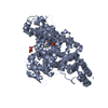
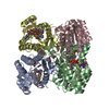
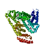
- PDB-3vpz: Crystal structure of glucokinase from Antarctic psychrotroph at 1.69A -
+ Open data

- Basic information
| Entry | Database: PDB / ID: 3vpz | ||||||
|---|---|---|---|---|---|---|---|
| Title | Crystal structure of glucokinase from Antarctic psychrotroph at 1.69A | ||||||
 Components Components | Glucokinase | ||||||
 Keywords Keywords | TRANSFERASE / GLUCOKINASE / COLD-ADAPTED | ||||||
| Function / homology |  Function and homology information Function and homology informationglucokinase / glucokinase activity / D-glucose binding / glycolytic process / ATP binding / cytosol Similarity search - Function | ||||||
| Biological species |  Pseudoalteromonas (bacteria) Pseudoalteromonas (bacteria) | ||||||
| Method |  X-RAY DIFFRACTION / SYNCHROTRON / MOLECULAR REPLACEMENT / Resolution: 1.69 Å X-RAY DIFFRACTION / SYNCHROTRON / MOLECULAR REPLACEMENT / Resolution: 1.69 Å | ||||||
 Authors Authors | Oda, T. / Fuchita, N. / Motoshima, H. / Kawamoto, M. / Watanabe, K. | ||||||
 Citation Citation | Journal: To be Published Title: Crystal structure of glucokinase from Antarctic psychrotroph at 1.69A Authors: Oda, T. / Fuchita, N. / Motoshima, H. / Kawamoto, M. / Watanabe, K. | ||||||
| History |
|
- Structure visualization
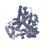
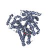
| Structure viewer | Molecule: MolmilJmol/JSmol |
|---|
- Downloads & links
-Download
| PDBx/mmCIF format | 3vpz.cif.gz | 77.6 KB | Display | PDBx/mmCIF format |
|---|---|---|---|---|
| PDB format | pdb3vpz.ent.gz | 57.5 KB | Display | PDB format |
| PDBx/mmJSON format | 3vpz.json.gz | Tree view | PDBx/mmJSON format | |
| Others |  Other downloads Other downloads |
-Validation report
| Arichive directory | https://data.pdbj.org/pub/pdb/validation_reports/vp/3vpzftp://data.pdbj.org/pub/pdb/validation_reports/vp/3vpz | HTTPS FTP |
|---|
-Related structure data
| Related structure data | 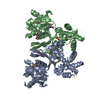 1q18S S: Starting model for refinement |
|---|---|
| Similar structure data |
-Links
 PDBj
PDBj- Assembly
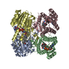
| Deposited unit | 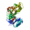
| ||||||||
|---|---|---|---|---|---|---|---|---|---|
| 1 | 
| ||||||||
| Unit cell |
|
-Components
| #1: Protein | Mass: 35131.703 Da / Num. of mol.: 1 Source method: isolated from a genetically manipulated source Source: (gene. exp.) Pseudoalteromonas (bacteria) / Strain: AS-131 / Gene: glk / Production host: |
|---|---|
| #2: Water | ChemComp-HOH /  Mass: 18.015 Da / Num. of mol.: 223 / Source method: isolated from a natural source / Formula: H2O Mass: 18.015 Da / Num. of mol.: 223 / Source method: isolated from a natural source / Formula: H2O |
| Has protein modification | Y |
-Experimental details
-Experiment
| Experiment | Method: X-RAY DIFFRACTION / Number of used crystals: 1 |
|---|
- Sample preparation
| Crystal | Density Matthews: 2.39 Å3/Da / Density % sol: 48.45 % |
|---|---|
| Crystal grow | Temperature: 293 K / Method: vapor diffusion, hanging drop / pH: 8.5 Details: 0.2M Trimethylamine N-oxide dihydrate, 20% PEGMME 2000, pH 8.5, VAPOR DIFFUSION, HANGING DROP, temperature 293K |
-Data collection
| Diffraction | Mean temperature: 100 K |
|---|---|
| Diffraction source | Source: SYNCHROTRON / Site: SAGA-LS  / Beamline: BL07 / Wavelength: 1.1 Å / Beamline: BL07 / Wavelength: 1.1 Å |
| Detector | Type: RIGAKU SATURN A200 / Detector: CCD / Date: Aug 31, 2011 |
| Radiation | Monochromator: Si(111) DOUBLE CRYSTAL / Protocol: SINGLE WAVELENGTH / Monochromatic (M) / Laue (L): M / Scattering type: x-ray |
| Radiation wavelength | Wavelength: 1.1 Å / Relative weight: 1 |
| Reflection | Resolution: 1.69→19.39 Å / Num. obs: 36190 / % possible obs: 95.8 % / Rmerge(I) obs: 0.075 / Net I/σ(I): 14.3 |
| Reflection shell | Highest resolution: 1.69 Å / % possible all: 95.8 |
- Processing
| Software |
| ||||||||||||||||||||||||||||||||||||||||||||||||||||||||||||||||||||||||||||||||||||||||||||||||||||||||||||||||||||||||||||||||||||||||||||||||||||||||||||||||||||||||||
|---|---|---|---|---|---|---|---|---|---|---|---|---|---|---|---|---|---|---|---|---|---|---|---|---|---|---|---|---|---|---|---|---|---|---|---|---|---|---|---|---|---|---|---|---|---|---|---|---|---|---|---|---|---|---|---|---|---|---|---|---|---|---|---|---|---|---|---|---|---|---|---|---|---|---|---|---|---|---|---|---|---|---|---|---|---|---|---|---|---|---|---|---|---|---|---|---|---|---|---|---|---|---|---|---|---|---|---|---|---|---|---|---|---|---|---|---|---|---|---|---|---|---|---|---|---|---|---|---|---|---|---|---|---|---|---|---|---|---|---|---|---|---|---|---|---|---|---|---|---|---|---|---|---|---|---|---|---|---|---|---|---|---|---|---|---|---|---|---|---|---|---|
| Refinement | Method to determine structure: MOLECULAR REPLACEMENT Starting model: PDB ENTRY 1Q18 Resolution: 1.69→18.56 Å / Cor.coef. Fo:Fc: 0.964 / Cor.coef. Fo:Fc free: 0.944 / SU B: 2.54 / SU ML: 0.084 / Isotropic thermal model: Isotropic / Cross valid method: THROUGHOUT / ESU R: 0.117 / ESU R Free: 0.12 / Stereochemistry target values: MAXIMUM LIKELIHOOD / Details: HYDROGENS HAVE BEEN USED IF PRESENT IN THE INPUT
| ||||||||||||||||||||||||||||||||||||||||||||||||||||||||||||||||||||||||||||||||||||||||||||||||||||||||||||||||||||||||||||||||||||||||||||||||||||||||||||||||||||||||||
| Solvent computation | Ion probe radii: 0.8 Å / Shrinkage radii: 0.8 Å / VDW probe radii: 1.2 Å / Solvent model: MASK | ||||||||||||||||||||||||||||||||||||||||||||||||||||||||||||||||||||||||||||||||||||||||||||||||||||||||||||||||||||||||||||||||||||||||||||||||||||||||||||||||||||||||||
| Displacement parameters | Biso mean: 37.076 Å2
| ||||||||||||||||||||||||||||||||||||||||||||||||||||||||||||||||||||||||||||||||||||||||||||||||||||||||||||||||||||||||||||||||||||||||||||||||||||||||||||||||||||||||||
| Refinement step | Cycle: LAST / Resolution: 1.69→18.56 Å
| ||||||||||||||||||||||||||||||||||||||||||||||||||||||||||||||||||||||||||||||||||||||||||||||||||||||||||||||||||||||||||||||||||||||||||||||||||||||||||||||||||||||||||
| Refine LS restraints |
| ||||||||||||||||||||||||||||||||||||||||||||||||||||||||||||||||||||||||||||||||||||||||||||||||||||||||||||||||||||||||||||||||||||||||||||||||||||||||||||||||||||||||||
| LS refinement shell | Resolution: 1.69→1.734 Å / Total num. of bins used: 20
|
