 Movie
Movie Controller

[English] 日本語
 Yorodumi
- PDB-3vnx: Crystal structure of ferritin from multicellular green algae, Ulv... -
+ Open data

- Basic information
| Entry | Database: PDB / ID: 3vnx | ||||||
|---|---|---|---|---|---|---|---|
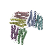
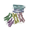
| Title | Crystal structure of ferritin from multicellular green algae, Ulva pertusa. | ||||||
 Components Components | ferritin | ||||||
 Keywords Keywords | OXIDOREDUCTASE / 4-helix bundle / Iron storage | ||||||
| Function / homology |  Function and homology information Function and homology informationferroxidase / ferroxidase activity / ferric iron binding / iron ion transport / ferrous iron binding / intracellular iron ion homeostasis / cytoplasm Similarity search - Function | ||||||
| Biological species |  Ulva pertusa (plant) Ulva pertusa (plant) | ||||||
| Method |  X-RAY DIFFRACTION / SYNCHROTRON / MOLECULAR REPLACEMENT / Resolution: 2.4 Å X-RAY DIFFRACTION / SYNCHROTRON / MOLECULAR REPLACEMENT / Resolution: 2.4 Å | ||||||
 Authors Authors | Masuda, T. / Morimoto, S.I. / Mikami, B. / Toyohara, H. | ||||||
 Citation Citation | Journal: Protein Sci. / Year: 2012 Title: The extension peptide of plant ferritin from sea lettuce contributes to shell stability and surface hydrophobicity. Authors: Masuda, T. / Morimoto, S.I. / Mikami, B. / Toyohara, H. | ||||||
| History |
|
- Structure visualization
| Structure viewer | Molecule: MolmilJmol/JSmol |
|---|
- Downloads & links
-Download
| PDBx/mmCIF format | 3vnx.cif.gz | 53.4 KB | Display | PDBx/mmCIF format |
|---|---|---|---|---|
| PDB format | pdb3vnx.ent.gz | 39.4 KB | Display | PDB format |
| PDBx/mmJSON format | 3vnx.json.gz | Tree view | PDBx/mmJSON format | |
| Others |  Other downloads Other downloads |
-Validation report
| Summary document | 3vnx_validation.pdf.gz | 427.9 KB | Display | wwPDB validaton report |
|---|---|---|---|---|
| Full document | 3vnx_full_validation.pdf.gz | 429.6 KB | Display | |
| Data in XML | 3vnx_validation.xml.gz | 9.8 KB | Display | |
| Data in CIF | 3vnx_validation.cif.gz | 13 KB | Display | |
| Arichive directory | https://data.pdbj.org/pub/pdb/validation_reports/vn/3vnxftp://data.pdbj.org/pub/pdb/validation_reports/vn/3vnx | HTTPS FTP |
-Related structure data
| Similar structure data |
|---|


-Links
 PDBj
PDBj


- Assembly
| Deposited unit | 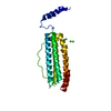
| |||||||||
|---|---|---|---|---|---|---|---|---|---|---|
| 1 | x 24
| |||||||||
| Unit cell |
| |||||||||
| Components on special symmetry positions |
|
-Components
| #1: Protein | Mass: 22877.609 Da / Num. of mol.: 1 Source method: isolated from a genetically manipulated source Source: (gene. exp.) Ulva pertusa (plant) / Plasmid: pET21d / Production host:  | ||||
|---|---|---|---|---|---|
| #2: Chemical | ChemComp-CA /   Mass: 40.078 Da / Num. of mol.: 4 / Source method: obtained synthetically / Formula: Ca Mass: 40.078 Da / Num. of mol.: 4 / Source method: obtained synthetically / Formula: Ca#3: Water | ChemComp-HOH / |  Mass: 18.015 Da / Num. of mol.: 71 / Source method: isolated from a natural source / Formula: H2O Mass: 18.015 Da / Num. of mol.: 71 / Source method: isolated from a natural source / Formula: H2OSequence details | A SEQUENCE DATABASE REFERENCE FOR THIS PROTEIN DOES NOT CURRENTLY EXIST. | |
-Experimental details
-Experiment
| Experiment | Method: X-RAY DIFFRACTION / Number of used crystals: 1 |
|---|
- Sample preparation
| Crystal | Density Matthews: 3.19 Å3/Da / Density % sol: 61.44 % |
|---|---|
| Crystal grow | Temperature: 293 K / Method: vapor diffusion, hanging drop / pH: 9 Details: 21% PEG 400, 0.1M Bicine, pH 9.0, VAPOR DIFFUSION, HANGING DROP, temperature 293K |
-Data collection
| Diffraction | Mean temperature: 100 K |
|---|---|
| Diffraction source | Source: SYNCHROTRON / Site: SPring-8  / Beamline: BL38B1 / Wavelength: 0.9 Å / Beamline: BL38B1 / Wavelength: 0.9 Å |
| Detector | Type: ADSC QUANTUM 315 / Detector: CCD / Date: Jun 4, 2011 |
| Radiation | Monochromator: double crystal monochromator / Protocol: SINGLE WAVELENGTH / Monochromatic (M) / Laue (L): M / Scattering type: x-ray |
| Radiation wavelength | Wavelength: 0.9 Å / Relative weight: 1 |
| Reflection | Resolution: 2.4→50 Å / Num. all: 22908 / Num. obs: 22816 / % possible obs: 99.6 % / Observed criterion σ(F): 2 / Observed criterion σ(I): 2 / Redundancy: 9.6 % / Biso Wilson estimate: 42.64 Å2 / Rmerge(I) obs: 0.055 / Net I/σ(I): 24.3 |
| Reflection shell | Resolution: 2.4→2.44 Å / Redundancy: 9.5 % / Rmerge(I) obs: 0.395 / Mean I/σ(I) obs: 9.21 / Num. unique all: 1122 / % possible all: 99.8 |
- Processing
| Software |
| ||||||||||||||||||||||||||||||
|---|---|---|---|---|---|---|---|---|---|---|---|---|---|---|---|---|---|---|---|---|---|---|---|---|---|---|---|---|---|---|---|
| Refinement | Method to determine structure: MOLECULAR REPLACEMENT / Resolution: 2.4→35.797 Å / SU ML: 0.24 / σ(F): 0.12 / Phase error: 21.45 / Stereochemistry target values: ML
| ||||||||||||||||||||||||||||||
| Solvent computation | Shrinkage radii: 0.9 Å / VDW probe radii: 1.11 Å / Solvent model: FLAT BULK SOLVENT MODEL / Bsol: 46.2 Å2 / ksol: 0.327 e/Å3 | ||||||||||||||||||||||||||||||
| Displacement parameters |
| ||||||||||||||||||||||||||||||
| Refine analyze | Luzzati coordinate error obs: 0.3336 Å | ||||||||||||||||||||||||||||||
| Refinement step | Cycle: LAST / Resolution: 2.4→35.797 Å
| ||||||||||||||||||||||||||||||
| Refine LS restraints |
| ||||||||||||||||||||||||||||||
| LS refinement shell | Refine-ID: X-RAY DIFFRACTION / Total num. of bins used: 4
|
